4574
Determination of the decrease rate of the apparent diffusion coefficient in the fetal kidneys during the third trimester.
LORENA AZYADE MURRIETA GONZALEZ1, carla maria garcia moreno2, and MARIA BARRERA ESPARZA3
1RESONANCIA MAGNETICA, HOSPITAL ANGELES LOMAS, Huixquilucan Estado de Mexico, Mexico, 2RESONANCIA MAGNETICA, Hospital Angeles Lomas, Huixquilucan, Mexico, 3RESONANCIA MAGNETICA, HOSPITAL ANGELES LOMAS, Huixquilucan, Mexico
1RESONANCIA MAGNETICA, HOSPITAL ANGELES LOMAS, Huixquilucan Estado de Mexico, Mexico, 2RESONANCIA MAGNETICA, Hospital Angeles Lomas, Huixquilucan, Mexico, 3RESONANCIA MAGNETICA, HOSPITAL ANGELES LOMAS, Huixquilucan, Mexico
Synopsis
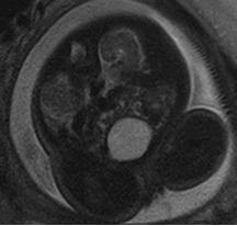
The main objective of the work was to determine the decrease rate of the apparent diffusion coefficient (ADC) in fetal kidneys using diffusion-weighted imaging (DWI) during the third trimester. We measured the renal ADC in 16 patients, and our findings exhibit an ADC decrease during the gestational age.
INTRODUCTION:
There are studies
that have shown a decrease of the ADC value in the fetal kidneys as the
gestational age increases. This has been related to the normal renal development.
Normal kidneys have a gradual increase in the number of glomeruli between the 10th
and 18th weeks of gestation, accelerating until the 32nd week. The renal perfusion is about 20 ml/min at 25
weeks gestation reaching to 60 ml/min at the end of gestation. 1.
Renal function has been assessed by indirect signs
such as the volume of amniotic fluid, the degree of pulmonary maturation and
the degree of bladder repletion; however, more recently, diffusion sequences (DWI)
have begun to be used as a tool to predict renal function 5,6.
DWI is considered the only method to assess molecular
diffusion in vivo. The apparent diffusion coefficient (ADC) values of the
renal parenchyma depends on Brownian movement, perfusion in the capillary
network and tubular flow (1).. Recently, MRI has begun to
demonstrate its value for obtaining additional findings in patients with
urinary system disorders that are not detected by ultrasound which result in a change
in postnatal management.1,2, 3, 4.
Some authors have demonstrated the usefulness of
diffusion for the detection ADC values change as gestational age progresses,
demonstrating the potential of magnetic resonance to detect developmental
variants. 7,8. The evaluation of renal functionality together with
lung maturity contributes to establishing the prognosis of the fetus.1, 8.
The values of the ADC of the fetal kidneys promises
to be a good way to predict the functionality, however, the rate of decrease of
the ADC during the third trimester has not yet been established. For these
reasons, the present study aims to know the values of the normal diffusion
coefficient of fetal kidneys in our population, as well as estimate the decrease
rate of ADC during the third trimester.
METHODS:
The images
we used were obtained at the MRI Department of the Ángeles Lomas Hospital, in México
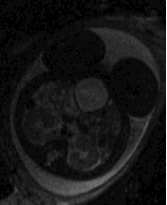
from July to November 2019. We used a 1.5T scanner (Siemens Aera) with a phased
arrayed coil placed over the mother´s pelvis. A DWI technique was used with b
values of 700 and 1000 s/mm2 and 12 directions. We studied 16
patients that were referred for pathology outside the kidneys.
We processed
the images using 3 different software: dcm2nii, to convert DICOM (digital imaging
and communication on medicine) images to NifTI (neuroimaging informatics
technology initiative) format and extract the gradient directions of diffusion;
FSL (functional magnetic resonance imaging of the brain software library),
where we correct eddy currents and movement effects, in addition to solve the
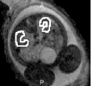
diffusion tensor and obtain the ADC for every voxel; and Image J, where
subsequently, the ADC mean value was obtained by performing a region of
interest (ROI) in axial slides covering the entire fetal renal parenchyma
avoiding the renal pelvis. Then we compared the ADC values corresponding to
each gestational age. Based on the previous studies, we assumed that the ADC
values have a linear decrease behavior with gestational age, so we show in the
results section the linear equations of the graphs with the ADC rate of
decrease.
RESULTS:
We found a correlation between fetal
kidneys ADC values and gestational age. ADC
values decrease with gestational age. This values ranging from 31 to 36 weeks
and from 1.15 to 2.67 × 10⁻³mm2s−1, standard
deviation (SD) 0.24.
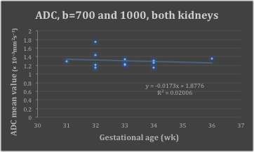
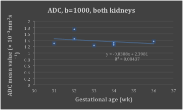
In Figures 1 and 2, we can observe
the behavior of ADC with gestational age, the equation that describe the curves
was obtained by linear regression. An analysis of Pearson's correlation
coefficient was performed between quantitative variables of ADC and gestational
age. In Figure 2, we obtained a decrease rate of 0.03 × 10⁻³mm2s−1 /week.
DISCUSSION: As we described previously, our results coincide with the works previously published. The ADC values show a decrease behavior with gestational age in normal fetal kidneys. There was a decreasing performance of ADC regardless of the b values that were used. and of the number of patients that were studied. (Figures 1 and 2). In order to conclude a standard behavior of ADC with gestation age, we need to increase our sample. Our results suggest that we can assume an abnormal fetal renal function if we find an abnormal ADC. CONCLUSION: Normal fetal kidneys have a decrease rate of 0.03 × 10⁻³mm2s−1 per week during the third trimester. This may provide important information related to renal function and development. Currently we are including more patients to increase the sample and determine a more precise standard behavior of ADC with gestational age in normal fetal kidneys. In the future we intend to study abnormal kideney´s ADC and compare them to gestational age paired controls.
DISCUSSION: As we described previously, our results coincide with the works previously published. The ADC values show a decrease behavior with gestational age in normal fetal kidneys. There was a decreasing performance of ADC regardless of the b values that were used. and of the number of patients that were studied. (Figures 1 and 2). In order to conclude a standard behavior of ADC with gestation age, we need to increase our sample. Our results suggest that we can assume an abnormal fetal renal function if we find an abnormal ADC. CONCLUSION: Normal fetal kidneys have a decrease rate of 0.03 × 10⁻³mm2s−1 per week during the third trimester. This may provide important information related to renal function and development. Currently we are including more patients to increase the sample and determine a more precise standard behavior of ADC with gestational age in normal fetal kidneys. In the future we intend to study abnormal kideney´s ADC and compare them to gestational age paired controls.
Acknowledgements
We would like to thank all radiological technical assistants for their practical support.References
- Manganaro L, Francioso A , et al . Fetal MRI with diffusion-weighted imaging (DWI) and apparent diffusion coefficient (ADC) assessment in the evaluation of renal development: preliminary experience in normal kidneys. Radiol. Med. 2009 Apr;114(3):403-13.
- Hörmann M, Brugger PC, et al. Fetal MRI of the urinary system. Eur J Radiol. 2006; 57:303-11.
- Gupta P, Kumar S, Sharma R, et al. The role of magnetic resonance imaging in fetal renal anomalies. Int J Gynaecol Obstet. 2010; 111:209-12.
- Prerna Gupta Sunesh Kumar, et al. El papel de la resonancia magnética en las anomalías renales fetales. Int J Gynaecol Obstet. Diciembre de 2010; 111 (3): 209-12.
- Savelli S, Di Maurizio M, et al. MRI with diffusion-weighted imaging (DWI) and apparent diffusion coefficient (ADC) assessment in the evaluation of normal and abnormal fetal kidneys: preliminary experience. Prenat Diagn. 2007; 27:1104-11.
- Chaumoitre K, Colavolpe N, et al. Diffusion-weighted magnetic resonance imaging with apparent diffusion coefficient (ADC) determination in normal and pathological fetal kidneys. Ultrasound Obstet Gynecol. 2007; 29:22-31.
- Witzani L 1, Brugger PC, et al. Normal renal development investigated with fetal MRI. Eur J Radiol. 2006 Feb;57(2):294-302.
- M. Gómez Huertas, M. Culianez Casas, et al. Papel complementario de la resonancia magnética en el estudio del sistema urinario fetal, European journal of radiology 2010;111:209-12.
