4573
ZOOMit DWI of the Fetal Brain: A Preliminary Evaluation Comparing DWI at 3T1BGI Company, ShenZhen, China, 2MR Collaborations, Siemens Healthcare Ltd, Shenzhen, China, 3Shenzhen Longgang District Central Hosptial, Shenzhen, China, 4Shenzhen Institutes of Advanced Technology, Shenzhen, China
Synopsis
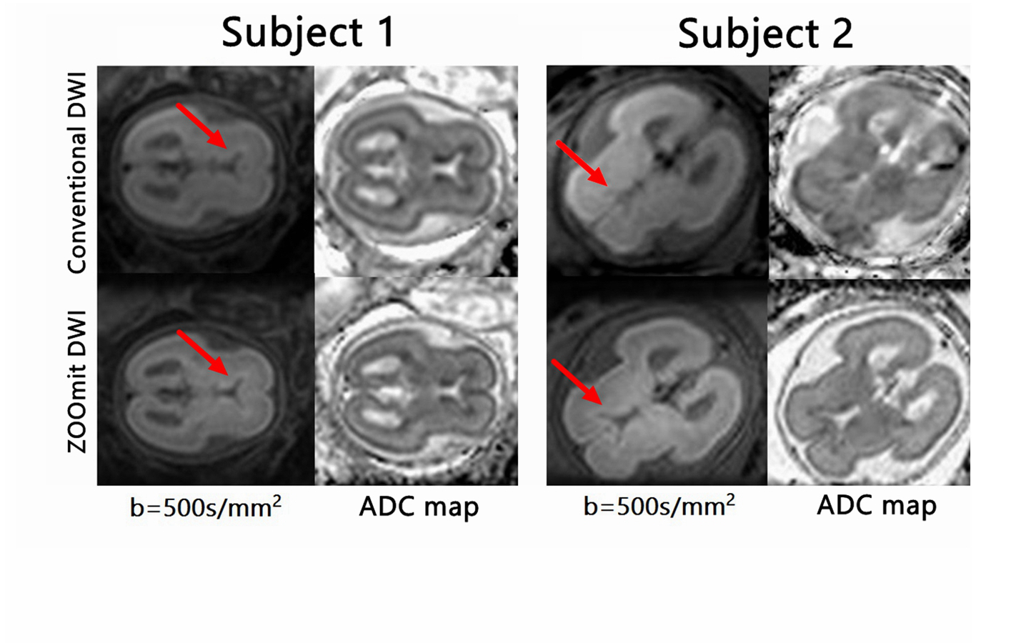
This study evaluated the clinical utility of zoomed diffusion-weighted echo-planar imaging (ZOOMit DWI) of the fetal brain. The effectiveness of the method was compared with conventional echo-planar DWI. Based on imaging analysis of 10 cases, the results showed that ZOOMit DWI exhibits markedly reduced susceptibility and distortion artifacts relative to conventional DWI. ZOOMit-DWI may serve as a viable alternative for the fetal brain assessment and pathology diagnosis.
Purpose
Echo-planar diffusion-weighted imaging (DWI) is increasingly performed to assess brain development in fetuses that are at increased risk for neurodevelopmental abnormalities. The method has a short acquisition time. However, the use of conventional echo-planar DWI for fetal brain imaging in pregnant patients is challenging due to chemical shift and susceptibility artifacts that may result in image ghosting and geometric distortion1-2. Zoomed diffusion-weighted EPI (ZOOMit DWI), by comparison, uses a combination of single-shot EPI with reduced-FOV imaging in the phase-encoding direction and spatially selective radiofrequency (RF) excitations. As such, this results in a decreased number of acquisition steps and EPI echo train reduction without any increase in scan time1-2. For these reasons, ZOOMit DWI may be useful to minimize the negative effects of the conventional method and, instead, provide improved-quality images. The aim of this study was to evaluate the feasibility and clinical robustness of a ZOOMit-DWI sequence in magnetic resonance imaging (MRI) of the fetal brain and compare the method with a conventional single-shot EPI sequence.Materials and Methods
This IRB-approved study included 10 participants (women with singleton pregnancies, 23-33 weeks gestation) who received imaging on a 3T MAGNETOM Prisma scanner (Siemens Healthcare, Erlangen, Germany) with an 18-channel body matrix coil. For conventional DWI, 3 b-values (50, 500, 800 s/mm2) were used, plus the following imaging parameters: TR/TE=3000/ 65ms; FOV=200x200 mm2; matrix=114×114; slice thickness=4mm, voxel size=1.8x1.8x4mm3, and slice number=15. For ZOOMit DWI, the same b-values were used along with the following imaging parameters: TR/TE=2800/57ms; FOV=190x87mm2; matrix=114×91; slice thickness=4 mm, voxel size=1.7x1.7x4mm3 and slice number=15. Scan time for the conventional DWI and ZOOMit DWI sequences were 1:32 minutes and 1:23 minutes, respectively. Signal-to-noise ratios (SNR) and apparent diffusion coefficient (ADC) values derived from the two methods were compared using the Wilcoxon signed-rank test. Two experienced radiologists scored the quality of the images generated by both two methods on a four-point scale (1 = poor, 4 = excellent). Statistical significance was defined as p < 0.05.Results
Compared with conventional DWI, ZOOMit DWI demonstrated less distortion and sharper depiction of the brain structures (Figure 1). The images obtained via the ZOOMit DWI method had an improved SNR (258.6±53.7 vs. 186.50±24.2, p=0.008). Furthermore, the ZOOMit DWI provided a superior image quality score compared to the conventional DWI (3.25±0.46 vs. 2.28±0.35, p=0.009). However, ADC values did not differ significantly between the two methods (1261±96 vs. 1253±89, p=0.20).Conclusion
Based on the results of this study, ZOOMit DWI of the fetal brain leads to substantial image quality improvements, exhibits markedly reduced susceptibility and distortion artifacts, plus shows higher spatial resolution of the wall relative to conventional DWI. The improved image quality obtained by ZOOMit DWI could potentially be helpful for the diagnosis of fetal brain pathologies. Additional clinical studies are recommended to validate the clinical value of the technique.Acknowledgements
No acknowledgement found.References
1. Schneider J F, Confort-Gouny S, Le Fur Y, et al. Diffusion-weighted imaging in normal fetal brain maturation[J]. European radiology, 2007, 17(9): 2422-2429.
2. Riffel P, Michaely H J, Morelli J N, et al. Zoomed EPI-DWI of the pancreas using two-dimensional spatially-selective radiofrequency excitation pulses[J]. PloS one, 2014, 9(3): e89468.
Figures