4572
Quantifying motion and temporal SNR in fetal functional MRI within a large cohort1Psychiatry, University of Michigan, Ann Arbor, MI, United States, 2Electrical Engineering and Computer Science, University of Michigan, Ann Arbor, MI, United States
Synopsis
In this work we leverage a large dataset consisting of 535 fMRI sessions from 248 unique subjects to assess the data quality of fetal resting state functional MRI. Our main goal is to describe the quality of fetal resting state fMRI after image post-processing. We quantify how much motion is present, apply a range of motion censoring thresholds to show how much data remains post-censoring, and evaluate the temporal signal to noise ratio of the time-series pre and post motion correction. Our results show very low average tSNR (2.7-3.7) and high percentages (40-95%) of discarded data.
Introduction
Fetal MRI is an exciting field that is experiencing rapid growth. There are numerous papers describing methods to improve aspects fetal MRI and fMRI, such as improved algorithms for spatiotemporal realignment (You 2016, Turk 2017) and improved imaging sequences (Ferazzi 2019). These methods have mostly focused on image acquisition and reconstruction techniques. There are few works that describe the characteristics of fetal fMRI post-processing. Rather than propose improved methods for processing fetal fMRI, in this work we aim to provide a detailed description of the data quality, assuming proper acquisition and post-processing occurred.Methods
Data were collected from 248 unique subjects. Some subjects were scanned longitudinally and contribute multiple sessions, thus the total number of MRI sessions in the present analysis is 535 (gestational age 20-37 weeks, M=30.9, SD=4.2). Data were acquired on a Siemens Verio 3T scanner using an abdominal 4-Channel Flex Coil (TR/TE: 2000/30; 4mm slice thickness, axial, interleaved ascending slice order). These data were preprocessed using an automated masking tool described in prior work (Rutherford 2019), which allows for the extraction of the fetal brain from maternal tissue across every volume within the time series. After segmenting the brain from other tissues and quality checking, time series were then realigned using MCFLIRT from FSL (middle volume as reference volume, 6 DOF, normcorr cost function). Realignment parameters output from MCFLIRT were then used to quantify the motion present using the framewise displacement (FD), a scalar quantity summarizing all 6 rigid body motion parameters, defined as: $$$FD_i = |\Delta d_{ix}| + | \Delta d_{iy}| + |\Delta d_{iz}| + |\Delta \alpha_i| + |\Delta \beta_i| + |\Delta \gamma_i|$$$, where $$$\Delta d_{ix} = d_{(i-1)x} - d_{ix}$$$. We then applied a range of framewise displacement motion censoring thresholds to determine the amounts of useable data remaining after censoring at each threshold. Next, we calculated the temporal SNR of the time series pre and post motion correction (realignment with no motion censoring). Temporal SNR is defined as the mean (across time) divided by the standard deviation (across time) of a voxel . We also investigated the relationships between fetal gestational age and temporal signal to noise ratio.Results
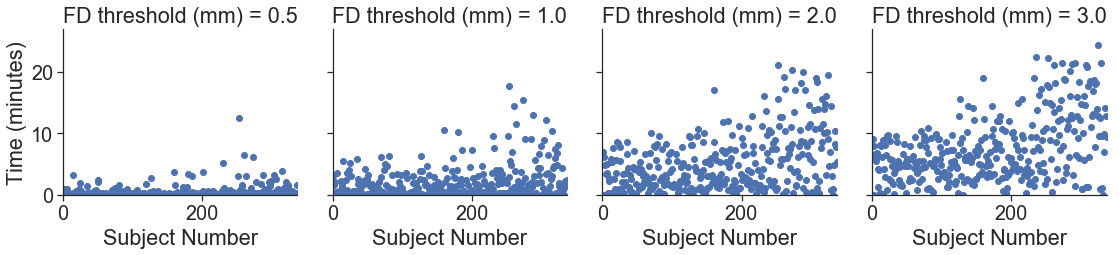
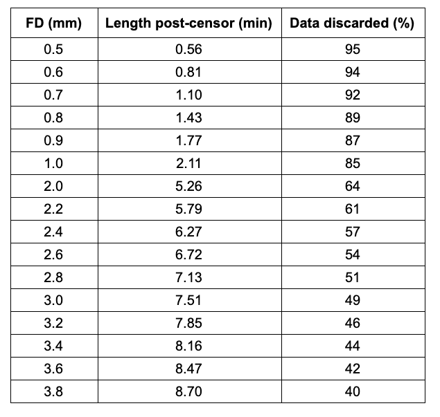
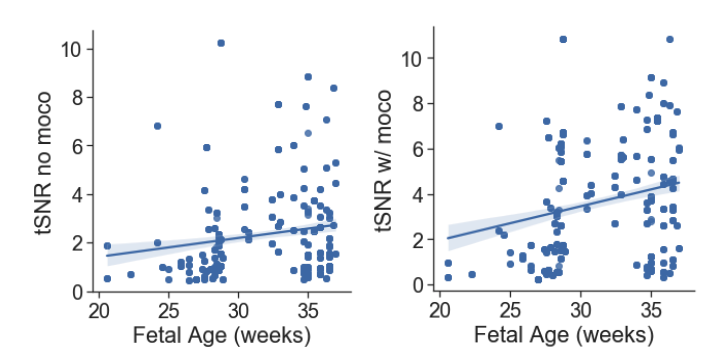
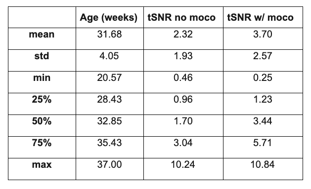
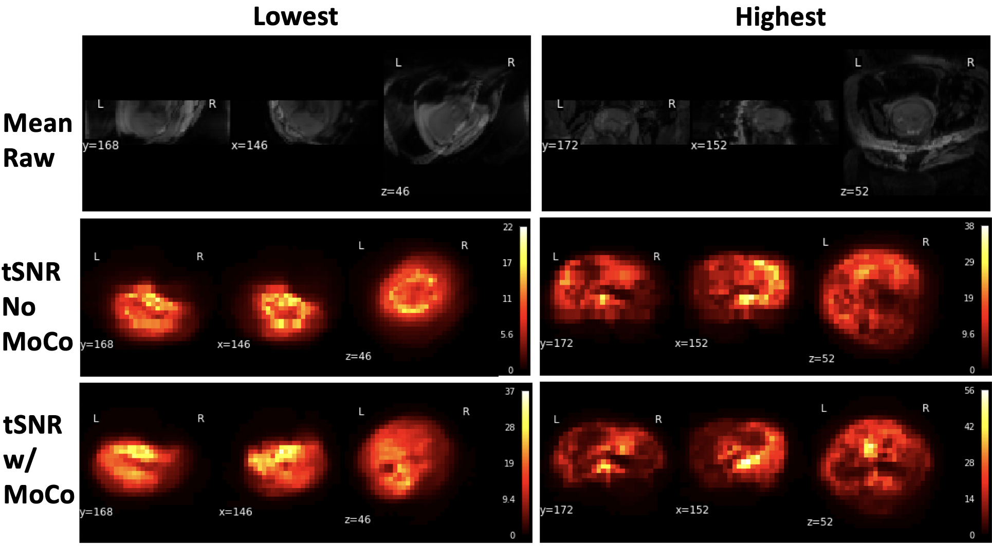
Fetal data exhibits substantial head motion. Figure 1 shows the length of scan after censoring for different threshold values calculated for each subject. For a threshold of FD>0.5mm, the scan was on average 0.56 minutes, resulting in 95% data loss. For the largest threshold of FD>3.8mm, the average scan length was 8.7 minutes, resulting in 40% data loss. Intermediate censoring threshold values are shown in Figure 2. The average temporal SNR of the segmented fetal brain prior to motion correction was 2.32 (sd=1.93) and the average tSNR post motion correction was 3.70 (sd=2.97). Both pre-motion correction tSNR (r=0.16, p=1.45e-3) and post-motion correction tSNR (r=0.24, p=3.54e-8) were significantly related to fetal gestational age (Figure 3, 4). Figure 5 shows example tSNR maps.Discussion & Conclusion
Fetal functional MRI is a challenging type of data to acquire with significant motion and lower tSNR than standard adult fMRI data. Nevertheless, there are an increasing number of researchers collecting these unique data. In this work we characterized the quality of our large sample of fetal resting state functional MRI data regarding motion (amount of data discarded and length of scan post censoring) and temporal signal to noise ratio. Depending on the motion censoring threshold used, anywhere between 40-95% of the fMRI scan will be discarded. Prior work in adult fMRI at 3T has shown that to detect a 1% signal change for 10 minutes of scanning requires a tSNR of 60 (Murphy, 2007). However, in fetal fMRI at 3T we found that with 15 minutes worth of scanning the average tSNR is only 2.32 (pre-motion correction) and 3.70 (post-motion correction)., This requirement of either long scanning with lower tSNR, or high tSNR scans for a shorter time, presents a large challenge for fetal fMRI data in detecting small changes in the BOLD signal, due to the large amounts of motion and lower tSNR. We invite further discussions regarding optimal motion censoring thresholds, gestational age at scan, and ideas for improving the future of fetal functional MRI.Acknowledgements
Thank you to the participant families who generously shared their time.References
Turk, E. A., Luo, J., Gagoski, B., Pascau, J., Bibbo, C., Robinson, J. N., Malpica, N. (2017). Spatiotemporal alignment of in utero BOLD-MRI series. Journal of magnetic resonance imaging : JMRI, 46(2), 403–412. doi:10.1002/jmri.25585
You, W., Evangelou, I. E., Zun, Z., Andescavage, N., & Limperopoulos, C. (2016). Robust preprocessing for stimulus-based functional MRI of the moving fetus. Journal of medical imaging (Bellingham, Wash.), 3(2), 026001. doi:10.1117/1.JMI.3.2.026001
Ferrazzi, G., Price, A. N., Teixeira, R., Cordero-Grande, L., Hutter, J., Gomes, A., … Hajnal, J. V. (2018). An efficient sequence for fetal brain imaging at 3T with enhanced T1 contrast and motion robustness. Magnetic resonance in medicine, 80(1), 137–146. doi:10.1002/mrm.27012
Rutherford, S., Sturmfels, P., Angstadt, M., Hect, J., Wiens, J., Van den Heuval, M., Scheinost, D., Thomason, M., Sripada, C. (2019). Automated Brain Masking of Fetal Functional MRI. bioRxiv 525386; doi:https://doi.org/10.1101/525386
Murphy, K., Bodurka, J., & Bandettini, P. A. (2007). How long to scan? The relationship between fMRI temporal signal to noise ratio and necessary scan duration. NeuroImage, 34(2), 565–574. doi:10.1016/j.neuroimage.2006.09.032
Figures