4568
Tracing and Therapeutic Evaluation of Transplanted Mesenchymal Stem Cell in The Spinal Cord Injury of Beagles using Diffusion Tensor Imaging1Nanjing Drum Tower Hospital Clinical College of Nanjing Medical University, Nanjing, China, 2Clinical Science, Philips Healthcare, Shanghai, China, 3Nanjing Drum Tower Hospital, Nanjing, China, 4Southeast University, Nanjing, China, 5Radiology, Nanjing Drum Tower Hospital, Nanjing, China
Synopsis
The labelled SPIO of the transplanted MSCs were found in spinal cord segment of TSCI based on T1w and T2w images. The DTI can be of great value in the dynamic evaluation of morphological and neurological changes in beagles after TSCI. FA values are correlated with the impairment and repair of the fiber tracts of the spinal cord. The FA value can satisfactorily evaluate the completeness of the fiber tracts after SCI and the repair of them by stem cells. The combination of conventional MRI and DTI is of great clinical importance for the diagnosis and therapeutic evaluation of SCI.
Introduction
Traumatic spinal cord injury (TSCI) refers to the structural and functional impairment of the spinal cord caused by various mechanical factors and it may result in complete or incomplete dysfunction in motion, sensation and sphincter below the injury level.In developed countries, the incidence of SCI is 13.1~163.4 among per million cases, while in underdeveloped countries, the rate is between 13.0 and 220.0 [1]. Currently, no effective clinical treatment has been found. However, recent animal model experiments confirmed that stem cell transplantation can realize cell regeneration and functional recovery of impaired spinal cord [2]. In current study, we combined the diffusion tensor imaging (DTI) with conventional MRI to dynamically evaluate morphological and neurological function in Beagles after their traumatic spinal cord injuries. Meanwhile, the current study investigates the in-vivo MRI tracing of SPIO labeled in the transplanted mesenchymal stem cells (MSCs) and evaluates the therapeutic effect of transplanting MSCs on repairing the injured spinal cord.Methods
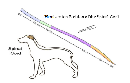
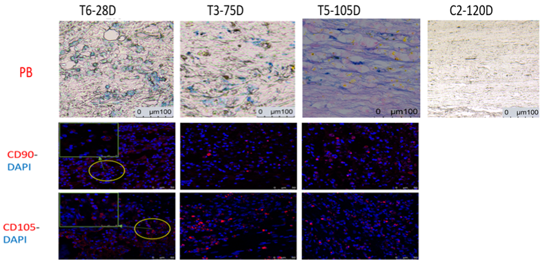
Three female beagles (age: 12-24 months, weight: 9-12 kg) are used to make an acute TSCI model by being performed a right hemisection of the spinal cord at T9 level (Figure 1). They are divided into three groups: group C (hemisection of the spinal cord, n=1), group M (hemisection of the spinal cord+ transplantation of huc-MSCs, n=1) and group T (hemisection of the spinal cord+ transplantation of Fe-huc-MSCs, n= 1). Multimodal MRI with T1w, T2w and DTI data are performed in 3.0T MRI scanner(Ingenia CX, Philips Healthcare, Best, The Netherland) and spinal BBB scores are conducted on these beagles respectively before and after the operation at an interval of 3d, 7d, 14d, 21d, 28d, 35d, 49d, 63d, and 77d. T1w and T2w images are used to perform the morphological evaluation of the injured spinal cord. The quantitative parameter FA calculated from DTI data can reflect the injury degree of the spinal cord and the repair of the injured spinal cord by MSCs, and the fiber tracts visually were traced using DTI-based tractography (DTT). At different timepoints, pathological and immunohistochemical examinations are performed and analyzed, including HE staining, PB staining, CD90+CD105 staining and CD68 staining, so as to verify if the MRI abnormal signals are related to the changes of iron signals with time, as well as to investigate migration, differentiation and other biological behaviors of the transplanted MSCs in the spinal cord.Results
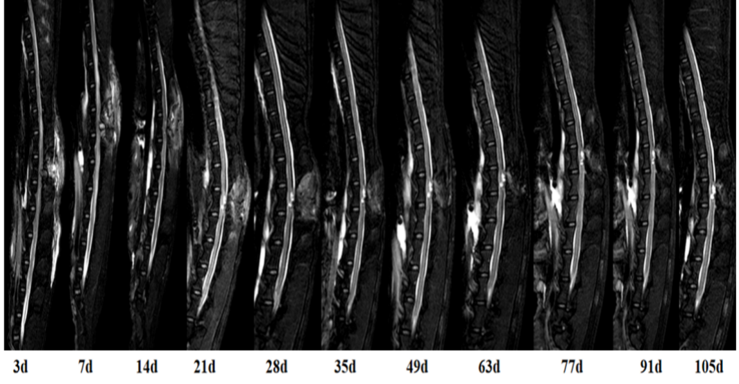
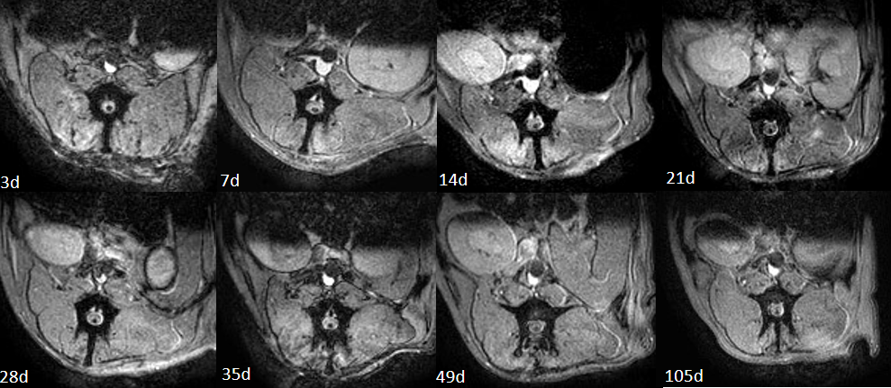
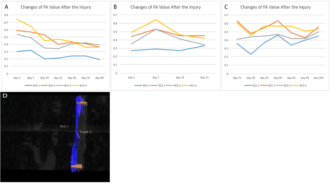
The spinal cord of these three beagles all undergoes swelling after the acute TSCI.At 7 days after injury, the swelling in the injured spinal cord disappears basically. After that, it can be observed on the T2w images that the injured area gradually turns into a intramedullary cystic cavity and its area can expand over time, which is the most common outcome after SCI and the physical barrier to repairing spinal fiber tracts (Figure 3). The DTI analysis shows that the FA value of all regions of interest (ROIs) in group C decrease with time, while that of injured areas in group M and group T slowly increase with time (Figure 4). It can be observed from the DTT result that the spinal fiber tracts are broken and sparse(Figure 4). The BBB score shows that the motion and sensation function of the dog in group M and group T recovers with time, while that in group C is the opposite. Spots of hyper-intense signals can be observed in T1w and T2w images at various time points in group T (Figure 2 and 3). Combined with the pathological results, it can be confirmed that the hyper-intense signals are the labelled SPIO of the transplanted MSCs and it is traced as long as 105d in vivo. Further immunohistochemical examinations shows that the SPIO in group T is still in the living transplanted MSCs after 28 days of the operation, but MSCs have been dead or differentiated into neural stem cells after 75 days of the surgery.Conclusion
The labelled SPIO of the transplanted MSCs were found in injury spinal cord segment of TSCI based on T1w and T2w images. The DTI can be of great value in the dynamic evaluation of morphological and neurological changes in beagles after TSCI. FA values are correlated with the impairment and repair of the white matter fiber tracts of the spinal cord. The FA value can satisfactorily evaluate the completeness of the white matter fiber tracts after SCI and the repair of them by transplanting stem cells. The combination of conventional MRI and DTI is of great clinical importance for the diagnosis and therapeutic evaluation of SCI.Acknowledgements
No acknowledgement found.References
[1] Ryken T C , Hurlbert R J , Hadley M N , et al. The Acute Cardiopulmonary Management of Patients With Cervical Spinal Cord Injuries[J]. Neurosurgery, 2013, 72:84-92.
[2] Michailidou C, Marston L, Souza L H D, et al. A systematic review of the prevalence of musculoskeletal pain, back and low back pain in people with spinal cord injury[J]. Disability & Rehabilitation, 2014, 36(9):705-715.
Figures