4552
A longitudinal study on heterogeneity of diffusional parameters of spontaneously-hypertensive rats.
Jung-Sen Hsiao1, Hung-Yu Fu1, Pei-Lun Yu1, Sheng-Min Huang1, Kung-Chu Ho2, and Fu-Nien Wang1
1Biomedical Engineering and Envionmental Sciences, National Tsing-Hua University, Hsinchu City, Taiwan, 2Chang-Gung Memorial Hospital, Taoyuan City, Taiwan
1Biomedical Engineering and Envionmental Sciences, National Tsing-Hua University, Hsinchu City, Taiwan, 2Chang-Gung Memorial Hospital, Taoyuan City, Taiwan
Synopsis
Diffusion MRI has been regarded as an assessment in characterizing brain tissue integrity. However, the information of tissue heterogeneity is often overlooked. To investigate the feasibility of diffusional heterogeneity in gray matter through DKI derived diffusivities, six spontaneously-hypertensive rats (SHR) in different ages were scanned on a 7T small animal MR scanner. The differences of heterogeneity of diffusivities and kurtosis between different ages were revealed, which may imply its possibility for tissue characterization. Therefore, the diffusional heterogeneity could be suggested to be a potential image-based biomarker for evaluating tissue integrity.
Introduction
Diffusion kurtosis imaging (DKI) have been used to characterize tissue integrity by extracting the diffusivities from region of interest (ROI). The averaged values of diffusivities are usually investigated, and the information of tissue heterogeneity is therefore disregarded. In this study, we studied the diffusional heterogeneity on rat models. Previous report revealed that DKI had demonstrated its possibility in detecting changes in gray matter1. We used spontaneously-hypertensive rats (SHR) as our animal model, because the cortex and putamen of SHR has been shown their cellular imbalance in different ages2. Therefore, we performed the analysis of heterogeneity of diffusional parameters for tissue characterization in gray matter of SHR.Materials and Methods
Six spontaneously-hypertensive rats were scanned on a 7T MR Bruker Clinscan scanner. SHR were divided into three groups (early = 12-14 weeks, n=6; middle = 22-32 weeks, n=6; late = 45 weeks, n=4). All subjects were anesthetized with 1.5% isoflurane mixed with oxygen. Diffusion weighted images were obtained using Siemens multidirectional diffusion weighting (MDDW) sequence with 30 gradient directions and 6 b-values (0, 500, 1000, 1500, 2000, 2500 s/mm2). The parameters were as follows: TR/TE = 3000/32 ms, matrix size = 92 x 92, NEX = 4, slice thickness = 1.5 mm, FOV = 35 x 35 mm2. DKI parameters such as mean, axial, radial, fractional anisotropy of diffusivities and kurtosis were calculated as MD/AD/RD/FA and MK/AK/RK/FAk, respectively. The non-diffusion weighted image (B0) from each individual was corregistered to one rat as a reference template in each group by SPM12. Two ROIs in gray matter were analyzed including cerebral cortex (CT) and caudate putamen (CPu). Tissue heterogeneity of each ROI was quantified by coefficient of variance (CV, CV = standard deviation/mean) of all voxels inside. The mean value of ROI was also calculated for comparison. Two-tailed t-test was applied to compare the various diffusivity and kurtosis of SHR in different age. P-value < 0.05 or < 0.01 were regarded as significant difference statistically, denoted on figures with the symbol + and *, respectively.Results
To investigate difference in SHR with different ages in selected ROIs, the traditional mean value derived from diffusivities and kurtosis maps by DKI were shown in figure 1. The CV derived from diffusivities and kurtosis maps by DKI were shown in figure 2. In figure 1, it was noticed that the error bar in each group in CPu was different, and therefore the CV was calculated. In figure 2, CV demonstrated that the difference between each group were magnified and easier to be observed, especially in MD, AD, and RD. Table 1 summarized the statistical result of tissue characterization ability between traditional mean value and CV. The result of two approaches both showed no significant difference when comparing early with middle stage, and significant difference between middle and late stage. It is noticed that only CV showed significant change between early versus late stage.Discussion
SHR has been considered as the animal model for hypertension and vascular cognitive impairment (VCI)3. VCI may be attributed to dementia, and it is almost irreversible once cognitive decline is manifested4. The current study demonstrated that diffusional heterogeneity revealed the difference between SHR in different stages. It is noted in table 1 that the CV of DKI could reveal the diffusional heterogeneity changes in early stage compared to late stage, and it could not be detected from traditional averaged parameters. The heterogeneities in CPu in late stage SHR were considerably reduced, which may be attributed to degradation caused by aging. It may imply that there are lost information using traditional parameters and therefore heterogeneity could be useful to complement. Reducing of diffusional heterogeneity may be a potential biomarker for evaluating tissue integrity. Further investigation of diffusional heterogeneity is suggested to be applied on detection of neurodegenerative diseases.Conclusion
Diffusional heterogeneities were different in selected ROIs of SHR in different stages. Therefore, the diffusional heterogeneity is anticipated as a potential image-based biomarker using previous disregarded information for evaluating tissue integrity.Acknowledgements
We thank the instrument support from Center for Advanced Molecular Imaging and Translation, Chang-Gung Memorial Hospital, Linkou.References
- Jens H. J, Joseph A. H, Anita R, et al. Diffusional kurtosis imaging: the quantification of non-gaussian water diffusion by means of magnetic resonance imaging. Magn Reson Med. 2005 Jun;53(6):1432-40.
- Marie-Françoise R, Felix F, Stefan T. E, et al. Cortical and Putamen Age-Related Changes in the Microvessel Density and Astrocyte Deficiency in Spontaneously Hypertensive and Stroke-Prone Spontaneously Hypertensive Rats. Curr Neurovasc Res. 2009 Nov;6(4):279-87.
- Tayebati SK, Tomassoni D, and Amenta F. Spontaneously hypertensive rat as a model of vascular brain disorder: microanatomy, neurochemistry and behavior. J Neurol Sci. 2012 Nov 15;322(1-2):241-9.
- López-Gil X, Amat-Roldan I, Tudela R, et al. DWI and complex brain network analysis predicts vascular cognitive impairment in spontaneous hypertensive rats undergoing executive function tests. Front Aging Neurosci. 2014 Jul 23;6:167.
Figures
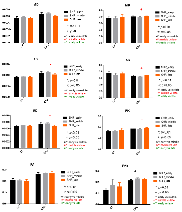
Figure 1. The traditional mean value derived from DKI.
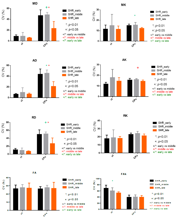
Figure 2. Diffusional heterogeneities of DKI were tested for the tissue characterization.
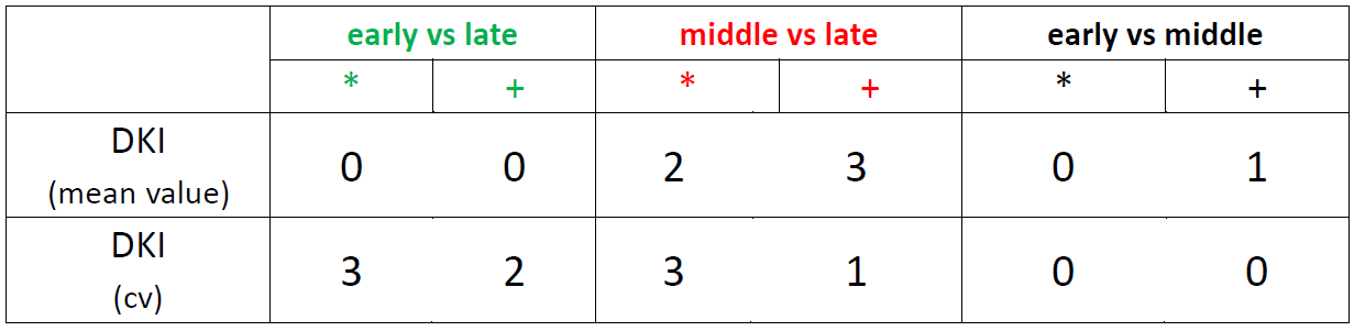
Table 1. Tissue characterization ability between mean value and CV by DKI. The number represents the total numbers in eight maps calculated from diffusivities and kurtosis, derived from DKI.