4492
Structural connectivity analysis of anterior nuclei of the thalamus in Papez circuit based on DW-MRI fiber tractography for ANT-DBS in epilepsy
Ruhunur Özdemir1,2, Kai Lehtimäki3, Jukka Peltola1,3, and Hannu Eskola1,2
1Department of Medicine and Health Technology, Tampere University, Tampere, Finland, 2Department of Radiology, Tampere University Hospital, Tampere, Finland, 3Department of Neuroscience and Rehabilitation, Tampere University Hospital, Tampere, Finland
1Department of Medicine and Health Technology, Tampere University, Tampere, Finland, 2Department of Radiology, Tampere University Hospital, Tampere, Finland, 3Department of Neuroscience and Rehabilitation, Tampere University Hospital, Tampere, Finland
Synopsis
The white matter organization of anterior nuclei of thalamus (ANT) has not been comprehensively examined before. We visualized white matter tracks in Papez circuit determining different ROIs in ANT, cingulate gyrus, mammillothalamic body, hippocampus and parahippocampal region for ANT-DBS in epilepsy. We employed constrained spherical deconvolution (CSD), multi-shell multi-tissue (MSMT) CSD, and DTI model to characterize white matter tissue organization in healthy controls and one patient diagnosed with refractory epilepsy in DW-MRI. We demonstrate a connectivity pattern of ANT. CSD model tractography may provide consistent insights on the alteration of the connections in patients with refractory epilepsy for performing ANT-DBS.
INTRODUCTION
Deep brain stimulation surgery (DBS) may provide an alternative treatment for the epileptic patients who are drug-resistant. Anterior nuclei of thalamus (ANT) has been a promising target in order to control and reduce epileptic seizures1. ANT is one of the member of the Papez circuit2. The clinical studies show that ANT presents different patterns of connectivity throughout the hippocampus, mammillary bodies, and neocortex3. However, the structural connection of ANT in Papez circuit has not been extensively examined. Diffusion MRI is a well-known technique that non‐invasively investigates the microstructural organization and orientation of biological tissues in vivo4. Diffusion tensor imaging (DTI) is one of the models to be widely accepted in order to examine the human brain, although it does not accurately perform in the presence of crossing fibres4. Constrained spherical deconvolution (CSD) has become one of widely used methods to extract full fibre orientations based on the estimation of fibre orientation distribution function overcoming the crossing fibre limitations5. Recent studies show that the b value and gradient directions also play a significant role in order to characterize white matter orientations in complex structures6; therefore, we also employed multi shell-multi tissue (MSMT-CSD)7 to achieve more reliable tractography. We aim to provide an insight into the structural connections of ANT and to demonstrate whether it is possible to detect any alteration in the epileptic brain.METHOD
We acquired high angular resolution DW-MRI data from five healthy volunteers and one patient with refractory epilepsy in 3T Siemens MAGNETOM Skyra MRI with a 64-channel head coil in 2.0×2.0×2.0 mm3 voxel size. The single shell scheme is 64 gradient directions at b= 3000 s/mm2 and multi shell scheme is 20, 30, 64 gradient directions respectively at b= 1000, 2000 ,3000 s/mm3 and four b= 0 images and also b=0 images were acquired with reverse-phase encoding to correct for the echo-planar imaging distortion correction and motion distortion. The other DW imaging parameters were Repetition time (TR) = 2900 ms, echo time (TE) = 120, The field of view, 260×260 s/mm2, the acquisition matrix, 130×130, and the number of excitations, 1. The number of the slice was 48 slices with no gap. High-resolution T1W MPRAGE was also acquired. The other parameters were TR 2000 ms, TE 2.45 ms, IT 960 ms, voxel size, 0.86 × 0.86 × 0.86 mm3. In the pre-processing pipeline, DW dataset were corrected for susceptibility-induced geometric distortions, eddy-current distortions, motion, and bias fields using FMRIB-FSL EDDY and TOPUP toolboxes. Three different tractography methods have been performed to generate the whole-brain connectome. First, we calculated tensors using eigenvectors in each voxel in order to perform DTI based tractography. Meanwhile, the estimations of response functions, finally the orientation distribution of the fibers in each voxel are computed in b value of 3000 s/mm3 to perform probabilistic CSD based tractography, and b values of 1000, 2000, 3000 s/mm3 to perform probabilistic MSMT-CSD based tractography using iFOD2 algorithm in MRtrix software. Whole-brain tractograms are generated using probabilistic DTI tracking, CSD tracking, MSMT-CSD. We filtered overestimated tracks in the whole-brain connectome in each individual. Ultimately, we visualized the structural connections determining ROIs in ANT, cingulate gyrus, mammillothalamic body, hippocampus, and parahippocampal region.RESULTS
We scanned five healthy controls (mean 35 years-of-age, 3 female and 2 male) and one patient (25 year-of-age) to provide an understanding of the structural connections of ANT and we aim to show whether it is possible to detect any alteration in the epileptic brain. We managed to demonstrate the white matter track organizations especially in ANT, mammillothalamic tract, cingulate bundle (anterior-posterior cingulate cortex) more accurately then the demonstration with DTI model.CONCLUSION
We visualized the connectomes of healthy controls and the patient to provide an insight on structural connectivity pattern of ANT in Papez circuit for ANT-DBS employing probabilistic DTI, MSMT-CSD and CSD tracking. The results show that probabilistic MSMT-CSD tracking is a more subtle approach to demonstrate the structural connectivity of ANT among these three approaches. In addition, this study will lead us to quantify the strength of the connections considering ROIs in healthy and epileptics individuals.Acknowledgements
The project has received funding from the European Union’s Horizon 2020 research and innovation program under the Marie Skłodowska-Curie grant agreement No 713645.References
- R.Fisher et al, Electrical stimulation of the anterior nucleus of thalamus for treatment of refractory epilepsy’, Epilepsia, March 2010, doi: 10.1111/j.1528-1167.2010.02536.x.
- Papez, J. W. , A proposed mechanism of emotion. Arch. Neurol. Psychiatry 38, 725–743, 1937, doi: 10.1001/archneurpsyc.1937.02260220069003
- Nicholas D. Child, Eduardo E. Benarroch, Anterior nucleus of the thalamus, functional organization and clinical implications, October 18, 2013, doi: https://doi.org/10.1212/01.wnl.0000436078.95856.56
- S. Mori et al, Principles of Diffusion Tensor Imaging and Its Applications to Basic Neuroscience Research, Neuron CellPress, Volume 51, Issue 5, 7 September 2006, Pages 527-539.
- J.D. Tournier et al, Robust determination of the fibre orientation distribution in diffusion MRI: Non-negativity constrained super-resolved spherical deconvolution, NeuroImage, Volume 35, Issue 4, 1 May 2007, Pages 1459-1472.
- Shawna Farquharson et al, White matter fiber tractography: why we need to move beyond DTI, Clinical article, J Neurosurg 118:1367–1377, 2013.
- B. Jeurissen, et al, Multi-tissue constrained spherical deconvolution for improved analysis of multi-shell diffusion MRI data. NeuroImage, 103 (2014), pp. 411–426
Figures
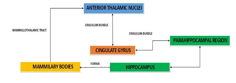
Schematic
diagram of ANT
in Papez circuit
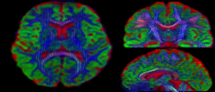
Estimation of Fiber Orientation Distribution (FOD) using MSMT-CSD
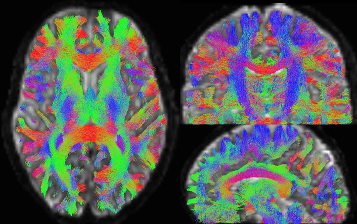
Connectome using probabilistic MSMT-CSD
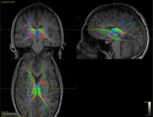
ROI: Anterior nucleus of thalamus using probabilistic MSMT-CSD
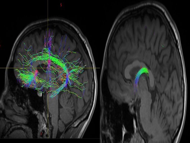
ROI: Cingulum Bundle and mammillothalamic tract using probabilistic MSMT-CSD