4370
Simultaneously multi-slice VAT-DIADEM at ultra-high field
Yi-Hang Tung1, Frank Godenschweger1, Myung-Ho In2, Alessandro Sciarra3, and Oliver Speck1
1Biomedical Magnetic Resonance, Otto-von-Guericke University Magdeburg, Magdeburg, Germany, 2Department of Radiology, Mayo Clinic, Rochester, MN, United States, 3Medicine and Digitalization, Otto-von-Guericke University Magdeburg, Magdeburg, Germany
1Biomedical Magnetic Resonance, Otto-von-Guericke University Magdeburg, Magdeburg, Germany, 2Department of Radiology, Mayo Clinic, Rochester, MN, United States, 3Medicine and Digitalization, Otto-von-Guericke University Magdeburg, Magdeburg, Germany
Synopsis
VAT-DIADEM is able to accelerate the distortion-free T2 weighting and diffusion weighting imaging sequence DIADEM at ultra-high field. The VAT gradient is turned on during imaging read-out in the slice selection direction. To further increase the acquisition efficiency, we developed a blipped version of the VAT gradient and applied simultaneous multi-slice imaging, based on blipped-CAIPI. The result is distortion reduction in EPI or acceleration in DIADEM and the increased volume coverage without increasing TR.
INTRODUCTION
A novel multi-shot EPI technique, termed DIADEM (Distortion-free Imaging: A Double Encoding Method)1,2 was proposed recently for distortion-free imaging. The DIADEM uses double encoding including the inherent EPI phase-encoding (EPI-PE) and additional spin-warp phase-encoding (SW-PE) to form a 3D data I(s,y,x). With this SW-PE, it can produce distortion-free and blurring-free T2-weighted and diffusion-weighted images. However, the extra loop of the SW-PE is time consuming. A common acceleration approach for DIADEM is the use of a reduced field of view (rFoV) in the SW-PE by skipping the step of SW-PE3. The unfolding process can be successful if the reduced FoV in the SW-PE direction still covers the entire range of geometric distortions. Otherwise, folding-over artifacts will appear in the calculated DIADEM image. A recent study4 demonstrated that further FoV reduction was achievable with view-angle-tilting (VAT) method by reducing the level of distortion in the EPI-PE direction.In this study, we propose the VAT-DIADEM combined with the simultaneous multi slice (SMS)5 acquisition to achieve higher acceleration factor in the acquisition and less folding-over ghost in the SMS-DIADEM data.
METHODS
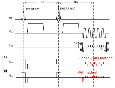
All the phantom scans were performed on a 7T scanner (Siemens Healthineers, Erlangen, Germany) using a 32-channel head coil (Nova Medical, Wilmington MA, USA). A SMS factor of 2 and no in-plane parallel imaging were applied to maximize the level of distortion and minimize the unfolding bias, which resulted in an echo-spacing of 1.0 ms. Other imaging parameters were: TR/TE = 1000.0/80.0 ms, 1.0×1.0×4.0 mm3 resolution with 300% distant factor, and the reduced resolution factor of 4 in EPI-PE. Figure 1 shows the sequence diagram with (a) SMS-DIADEM (b) SMS-VAT-DIADEM. The VAT gradient amplitude is set to the same as in the blipped-CAIPI gradient amplitude. As the SMS-DIADEM sequence is unfolded by single-band EPI reference, SMS-VAT-DIADEM is similarly unfolded by VAT-EPI.RESULTS and DISCUSSION
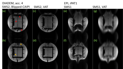
Figure 2 shows the unfolded SMS-EPI and DIADEM images with blipped-CAIPI gradients and blipped VAT gradients for the same acceleration (i.e. rFoV) factor of 4. From Figure 2 (e,f) SMS-EPI and (g,h) SMS-VAT-EPI it can be seen that unfolded EPI images show less distortion, but increased image blurring for VAT gradient. From Figure 2 (a,b) SMS-DIADEM and (c,d) SMS-VAT-DIADEM show that fold-over artifacts only appears in the SMS-DIADEM for an acceleration factor of 4. The highest fold-over-free rFoV acceleration factor is 2 for SMS-DIADEM and 4 for SMS-VAT-DIADEM. The value is consistent with previous VAT studies4,6. Although signal-to-noise ratio for SMS-VAT-DIADEM is around 40% of SMS-DIADEM, the differences could be minimized with a high VAT gradient amplitude and the use of a high in-plane parallel imaging factor4. Note that single-band images are in good consistence with multi-band images (data not shown).CONCLUSION
In this work, SMS is additionally added into the VAT-DIADEM to further accelerate the DIADEM acquisition. Combining both the VAT and SMS gradients on the slice axis allows rapid DIADEM acquisition not only in the in-plane (SW-PE) but also in the though-plane direction.Acknowledgements
This project is supported by DFG-grant SP 632/4-2.References
- In MH, et al., NeuroImage. 2017; 148:20-30 2
- In MH, et al., JMRI. 2019
- Zaitsev M et.al., MRM 2004
- Tung YH et al., ISMRM 2018, p.1024
- Setsompop K et al., MRM 2012
- Tung YH et al., ESMRMB 2019
Figures

Figure 1. SMS-VAT-DIADEM
sequence. (a) Conventional SMS slice
encoding gradients have blips in different polarities. (b) VAT SMS slice encoding gradients having blips with the same
polarity enable to reduce the distortion in EPI and
thus to further accelerate the DIADEM acquisition.

Figure 2. SMS-DIADEM vs. SMS-VAT-DIADEM: Results
with acceleration
(rFOV) factor of 4 in the SW-PE and SMS factor of 2 are shown. EPI (e, f) has severe distortion in the
EPI-PE direction, while VAT-EPI (g,
h) can reduce distortion, but at the expense of increased blurring. In conventional SMS-DIADEM (a, b), high acceleration in DIADEM causes fold-over artifacts due to strong EPI distortion. In
distinction, SMS-VAT-DIADEM
(c, d) allows a high acceleration factor of 4 in DIADEM acquisition without fold-over artifacts. Arrows: fold-over
artifacts or blurring artifacts.