4364
Highly Accelerated and High-Quality Intra-voxel Incoherent Motion DWI Enabled by Parametric POCS based multiplexed sensitivity-encoding1The University of Hong Kong, Hong Kong, Hong Kong, 2Graduate Institute of Biomedical Electronics and Bioinformatics, National Taiwan University, Taipei, Taiwan, Taipei, Taiwan, 3Department of Medical Imaging, China Medical University Hsinchu Hospital, Taiwan, Taipei, Taiwan
Synopsis
Low SNR and long acquisition time hinder the clinical feasibility of intra-voxel incoherent motion (IVIM) diffusion-weighted imaging. In this work, we used a joint reconstruction framework based on POCSMUSE algorithm to simultaneously reconstruct under-sampled multi-b multi-shot DW-EPI data without undesired noise amplification. A proposed parametric correction scheme (PCS) was incorporated into POCSMUSE algorithm for robust reconstruction based on either conventional bi-exponential or simplified IVIM model. The proposed method demonstrated that the high-quality brain IVIM images could be reconstructed from highly accelerated (R=4) data acquired with multi-b multi-shot DW-EPI.
Introduction
Intra-voxel incoherent motion (IVIM) diffusion-weighted imaging (DWI) is shown to be useful in assessing diffusion and perfusion characteristics of brain tissue1, 2. In practice, IVIM DWI utilizes single-shot diffusion-weighted echo-planar imaging (ssDW-EPI) to acquire a data set with multiple b-values1, 2, and subsequently derives the tissue parameters using a bi-exponential model. However, the use of ssDW-EPI leads to suboptimal data quality, such as in the presence of geometric distortion and limited spatial resolution. To improve the image quality, ssDW-EPI is combined with SENSE3 to reduce the distortion and improve the spatial resolution, but at the expense of noise amplification. Although multi-shot DW-EPI (msDW-EPI) with POCSMUSE reconstruction can obtain high-resolution and high-quality DWI data without undesired noise amplification4, the prolonged scan time due to multi-shot acquisition is impractical for the clinical acquisition of IVIM DWI. Recently, a parametric POCSMUSE framework has been reported to simultaneous generate high-quality multi-echo T2W images and accurate T2 maps from GRASE-EPI sequence with high scan acceleration5. In light of this, we proposed to improve the image reconstruction of highly accelerated multi-b msDW-EPI data, by incorporating a parametric correction scheme (PCS) into parametric POCSMUSE framework.Methods
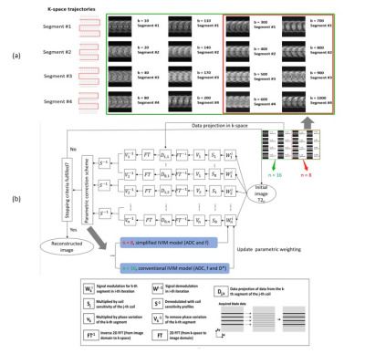
Data Acquisition and Hybrid-Simulation: All data were collected from three healthy volunteers on a 1.5T MRI scanner (GE Explore Lift) using a 12-channel head coil. For each subject, two brain IVIM datasets were acquired by using a 4-shot DW-EPI sequence with and without CSF-suppression, respectively. Seventeen b-values (0, 10, 20, 40, 80, 110, 140, 170, 200, 300, 400, 500, 600, 700, 800, 900, and 1000 s/mm2) with three orthogonal diffusion directions for each b-value were acquired for each IVIM dataset with the scan parameters shown as follow: TE/TR=84/4000ms, TI=1500ms, matrix size=128x128, slice thickness=5mm, and scantime=13min. After acquisition, the fully-sampled 4-shot DW-EPI data was used to simulate an under-sampled IVIM dataset with high acceleration factor (i.e., R=4), by selecting 1 out of 4 k-space segments for each multi-b data (Figure 1a).Image Reconstruction: Figure 1b shows the flowchart of parametric POCSMUSE algorithm with proposed PCS for simultaneously reconstructing highly accelerated multi-b images of a single diffusion direction. The fully-sampled b=0 image and corresponding ADC map (derived from fully-sampled b=0 and b=600 images) served as intimal data at the beginning of first iteration. After several iterations, either conventional bi-exponential or simplified IVIM model was applied in PCS to perform IVIM curve fitting. The new ADC, f, and D* derived from PSC at current iteration were used to update the diffusion-modulation operator for next iteration (n is the number of b-values). The reconstructed multi-b images were output from proposed framework when the iteration converged. Afterward, the image data with three orthogonal diffusion directions were averaged together for generated mean diffusion image for each b-value. In addition, the fully-sampled and under-sampled IVIM data were respectively reconstructed with MUSE and SENSE for comparison. The average multi-b DW images produced by three different reconstruction methods were then used for estimating IVIM parameters using either conventional bi-exponential IVIM model (with 16 b-values) or simplified model IVIM model (with 8 b-values).
Results
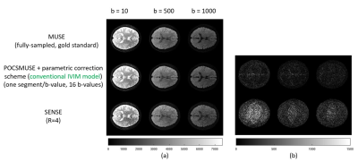
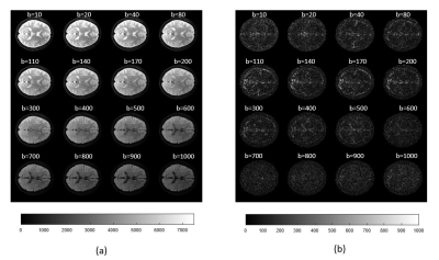
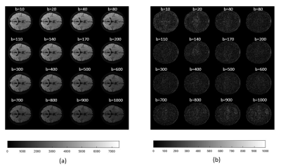
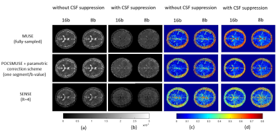
Figure 2a shows three representative images of 16 multi-b DW images reconstructed with MUSE, proposed framework, and SENSE for comparison. Figure 2b shows difference maps of reconstruction image between gold standard (fully-sampled multi-b DW images reconstructed using MUSE algorithm) and the other two reconstruction methods. Figures 3a and 4a show multi-b DW images reconstructed from the data with and without CSF-suppression, using proposed POCSMUSE framework with conventional bi-exponential IVIM model for PCS. Figures 3b and 4b show the difference maps of reconstructed image between gold standard (MUSE) and proposed framework. Figure 5 shows ADC maps and perfusion fraction maps derived from the multi-b DW images (with and without CSF suppression) reconstructed with three different methods.Discussion and Conclusion
Our proposed parametric POCSMUSE algorithm with PCS can successfully eliminate aliasing artifacts in highly under-sampled multi-b DW images without undesired noise amplification due to SENSE reconstruction. The IVIM parameter mapping results have demonstrated that the proposed framework is compatible with either conventional bi-exponential or simplified IVIM model, and can obtain quantitative maps close to the gold standards (4-shot fully-sampled data with MUSE). In addition to reduced noise and improved geometric fidelity, the proposed framework provides a substantial reduction in scantime compared to the data acquired with multi-shot (i.e., 27% of the scantime for acquiring fully-sampled data). Even though SENSE-produced multi-b DW images were aliasing-free and distortion-mitigated, the undesired noise amplification may affect the accuracy in the measurements of ADC and perfusion fraction (the bottom row in Figure 5). In the preliminary evaluation, the partial volume effect, such as the voxel involves both brain tissue and CSF signals, can lead to the bias in IVIM parameter estimation when using proposed framework. This limitation may be addressed by using CSF-suppression for data acquisition. Our results also demonstrate the feasibility of deriving IVIM parameter from CSF-suppressed data using proposed framework (Figures 3b vs 4b). In conclusion, the proposed framework can enable IVIM data acquisition with high acceleration factor, thereby improving the geometric fidelity and feasibility of IVIM for clinical application.Acknowledgements
The work was in part supported by grants from Hong Kong Research Grant Council (GRF HKU17138616 and GRF HKU17121517), and Hong Kong Innovation and Technology Commission (ITS/403/18).References
1. Suh, C.H., H.S. Kim, S.S. Lee, et al. Atypical imaging features of primary central nervous system lymphoma that mimics glioblastoma: utility of intravoxel incoherent motion MR imaging. Radiology. 2014; 272(2): 504-513.
2. Zhang, S.-x., Q.-j. Jia, Z.-p. Zhang, et al. Intravoxel incoherent motion MRI: emerging applications for nasopharyngeal carcinoma at the primary site. European radiology. 2014; 24(8): 1998-2004.
3. Pruessmann, K.P., M. Weiger, M.B. Scheidegger, and P. Boesiger. SENSE: sensitivity encoding for fast MRI. Magnetic resonance in medicine. 1999; 42(5): 952-962.
4. Chu, M.L., H.C. Chang, H.W. Chung, et al. POCS‐based reconstruction of multiplexed sensitivity encoded MRI (POCSMUSE): a general algorithm for reducing motion‐related artifacts. Magnetic resonance in medicine. 2015; 74(5): 1336-1348.
5. Chu, M.L., H.C. Chang, K. Oshio, and N.k. Chen. A single‐shot T2 mapping protocol based on echo‐split gradient‐spin‐echo acquisition and parametric multiplexed sensitivity encoding based on projection onto convex sets reconstruction. Magnetic resonance in medicine. 2018; 79(1): 383-393.
Figures
