4357
PROPELLER Diffusion-Weighted Imaging of the Prostate with Deep-Learning Reconstruction1Global MR Applications & Workflow, GE Healthcare, Houston, TX, United States, 2Global MR Applications & Workflow, GE Healthcare, New York, NY, United States, 3Global MR Applications & Workflow, GE Healthcare, Hino, Japan, 4Radiology, University of Wisconsin Madison, Madison, WI, United States, 5Global MR Applications & Workflow, GE Healthcare, Waukesha, WI, United States
Synopsis
While echo-planar diffusion-weighted imaging (EP-DWI) is the main sequence for cancer detection in the prostate peripheral zone, it is susceptible to signal loss and distortion due to B0-field inhomogeneities secondary to a variety of causes, including rectal gas or metal hardware in the pelvis. We were able to demonstrate that a spin-echo based DWI sequence with radial k-space sampling (PROPELLER) can overcome such artifacts and the addition of a deep-learning reconstruction algorithm can overcome the poor signal-to-noise (SNR) profile of the PROPELLER-DWI, overall generating images with minimal-to-no appreciable artifact and favorable SNR.
INTRODUCTION
Echo-planar diffusion-weighted imaging (EP-DWI) is the main sequence for lesion detection in the peripheral zone of the prostate, where the majority of cancers arise [1]. However, this sequence is susceptible to signal loss and distortion due to B0-field inhomogeneities secondary to a variety of causes, including rectal gas or metal hardware in the pelvis. While spin-echo based DWI sequences can improve susceptibility and metal-associated artifacts, they face noteworthy limitations, mainly a poor signal-to-noise (SNR) profile, which limits their utilization in prostate MRI where high b-value DWI is needed. Hence, we propose a novel DWI technique aimed to reduce susceptibility artifact by radial k-space sampling (PROPELLER) [2] and maintain comparable image acquisition times by fewer number of signal averages, compensated by a prototype deep-learning (DL) image reconstruction algorithm to reduce image noise.METHODS
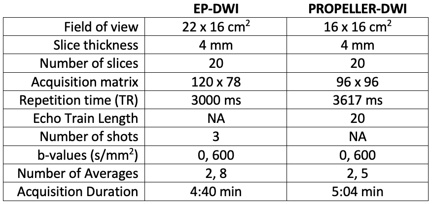
Imaging studies were performed on 3T MRI scanners (SIGNA Premier, GE Healthcare, Waukesha, USA), using a 16-channel anterior surface coil and a 60-channel posterior coil embedded within the table. Figure 1 includes a table summarizing the image acquisition parameters. Images were obtained on healthy volunteers. Two sets of images were generated from the PROPELLER-DWI raw data: one with conventional reconstruction (referred to as Conv-PROPELLER-DWI) and the other with a prototype DL reconstruction (referred to as DL-PROPELLER-DWI); the DL reconstruction algorithm was comprised of a deep convolutional residual encoder network trained using a prior database to improve SNR and reduce artifacts. Regions of interest (ROIs) were drawn on the peripheral zone and transition zone on the apparent diffusion coefficient (ADC) maps to evaluate the effect of different reconstruction algorithms on ADC measurements. Synthetic high b-value images at 1000 and 1500 s/mm2 were generated for all image sets.RESULTS
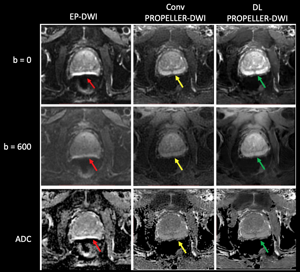
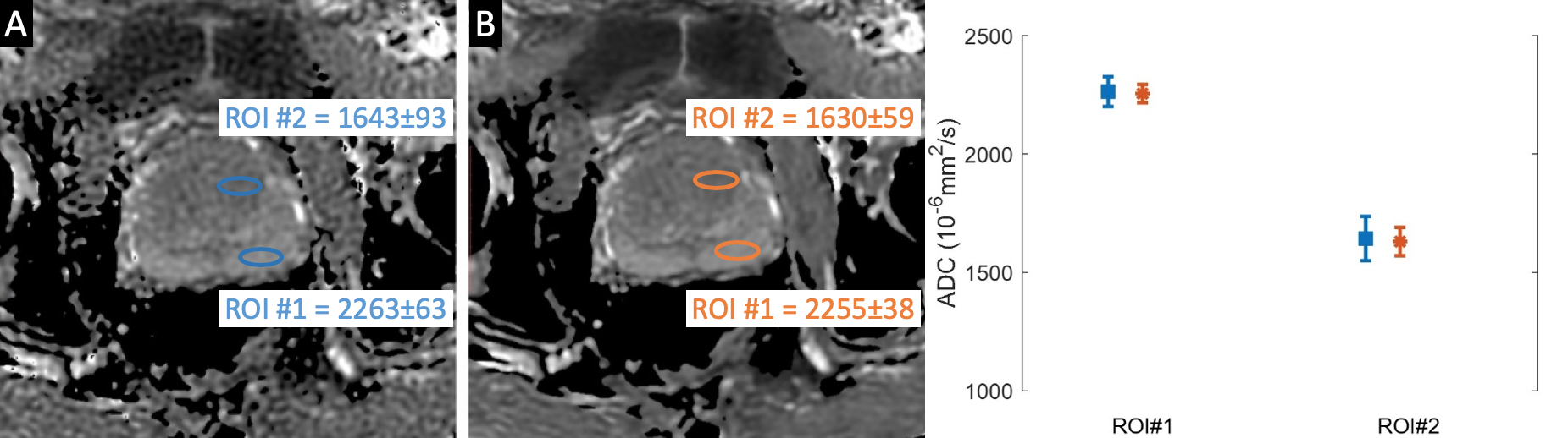
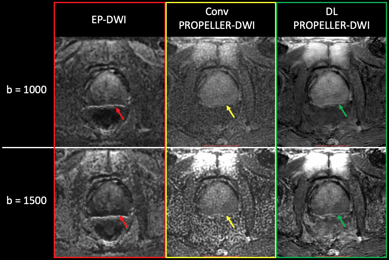
While EP-DWI images suffered significant artifact in the peripheral zone, Conv-PROPELLER-DWI was diagnostic and without appreciable artifact (Figure 2). While the subjective SNR was comparable between the EP-DWI and Conv-PROPELLER-DWI at b = 0 s/mm2, the latter suffered from a subjective yet appreciable decrease in SNR at b = 600 s/mm2. However, DL-PROPELLER-DWI ameliorated both the artifact seen on EP-DWI as well as the low SNR on Conv-PROPELLER-DWI, generating images without appreciable artifact and a favorable SNR. The same pattern is reflected in the ADC maps generated by each image set, with the DL-PROPELLER-ADC demonstrating the most favorable image quality.The ROI comparison between Conv- and DL-PROPELLER-DWI demonstrated similar ADC values, with a smaller standard deviation within the ROI on the DL-ADC map, as detailed in Figure 3. Synthetic high b-value images at 1000 and 1500 s/mm2 followed the same pattern as the acquired images at lower b-values (Figure 4); i.e., EP-DWI suffered from susceptibility artifact in the peripheral zone, rendering the images non-diagnostic, while Conv-PROPELLER-DWI did not exhibit the artifact, albiet with reduced SNR. DL-PROPELLER-DWI was able to ameliorate both the artifact and the SNR challenge, producing the best images overall.
DISCUSSION/CONCLUSIONS
Addition of DL image reconstruction to PROPELLER-DWI demonstrates an exciting potential to overcome the obstacles in the way of utilization of this method as a diagnostic tool for detection of prostate cancer. DL-PROPELLER-DWI benefits from both artifact reduction (compared to the current EP-DWI technique) and favorable SNR (compared to the conventional PROPELLER-DWI). While a formal evaluation is required to determine the lesion detectability of this technique, the preliminary results are indeed promising, and we are conducting the next steps in patients with, or at risk for, prostate cancer.Acknowledgements
No acknowledgement found.References
1. Weinreb JC, Barentsz JO, Choyke PL, et al. PI-RADS Prostate Imaging - Reporting and Data System: 2015, Version 2. Eur Urol 2016; 69:16-40
2. Czarniecki M, Caglic I, Grist JT, et al. Role of PROPELLER-DWI of the prostate in reducing distortion and artefact from total hip replacement metalwork. Eur J Radiol 2018; 102:213-219
Figures