4350
A low-rank based reconstruction method for diffusion-weighted point-spread-function encoded EPI (PSF-EPI)1Center for Biomedical Imaging Research, Department of Biomedical Engineering, School of Medicine, Tsinghua University, Beijing, China, 2Philips Healthcare, Beijing, China
Synopsis
Recently, a distortion- and blurring-free acquisition method called PSF-EPI has been used in DWI. However, when field inhomogeneity is severe, DW images may become noisy and more shots are needed to get the reliable results. In this work, we introduce a low-rank based reconstruction method using signal correlation along the ky-encoding dimension in PSF-EPI to improve image quality and reduce needed shot number to shorten scan time. High-resolution in-vivo data were used to test the performance of the proposed method. The results show that the quality of the images is improved.
Introduction
Diffusion-weighted imaging (DWI) plays a crucial role in clinical neurology and neuroscience studies1. Due to its fast acquisition speed, single-shot EPI is widely used in DWI. However, with limited bandwidth along the phase encoding direction, single-shot EPI suffers from distortion and blurring. Recently, efforts have been made to use multi-shot methods to address these problems2-4. Among them, a new point-spread-function (PSF) encoded EPI using Tilted-CAIPI acquisition and reconstruction has been proposed to provide distortion- and blurring-free images with acceleration factors over 20 fold5-6. Nevertheless, when field inhomogeneity is severe, it can be difficult to recover the undersampled data correctly and more shots are needed to improve reliability, which increases scan time. In this work, based on the similar low frequency content among images obtained at different ky locations in PSF-EPI, we developed a low-rank based method using the signal correlation along the ky-encoding dimension to improve the accuracy of reconstruction.Theory
PSF-EPIIn PSF-EPI, an additional phase-encoding called PSF-encoding is added before the original EPI readout. Since different PSF-encoding signals obtained at the same ky are acquired at the same echo times, PSF direction is a distortion-free dimension. Thus, 2D distortion- and blurring-free images acquired at different echo times can be obtained.
Low rank property for PSF-EPI
In PSF-EPI acquisition, a 2D(x-psf) image obtained at ky =m can be described as follows,
$$I_m=I_0 e^{-m∆t/T_2^* } e^{-ik_m y}e^{-iγ∆Bm∆t}$$
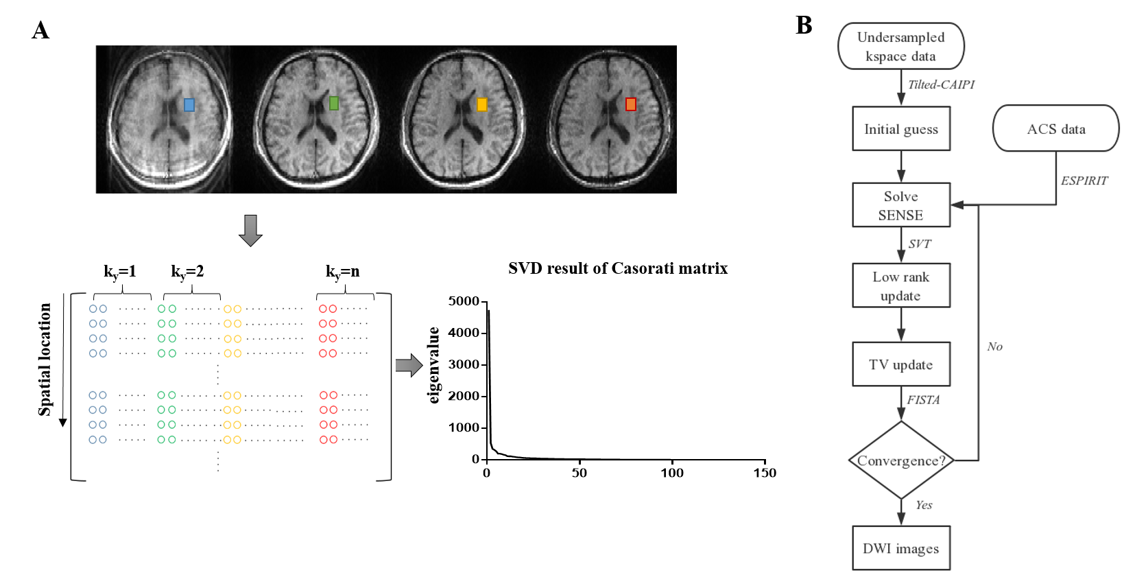
Since these images share the same low frequency magnitude component $$$I_0$$$, correlation exists between these 2D images and a low rank matrix can be formed. As shown in Figure. 1A, we construct the low rank matrix by stacking points at the same location from different x-psf images together. The different colors represent images at different ky locations. The SVD result proves its low rank property.
Reconstruction model
Considering that optimization for low rank constraint is a non-convex problem, the nucleus norm penalty is used. By minimizing the nucleus norm, the rank of the matrix will be reduced. This optimization is achieved by single value thresholding (SVT) algorithm.
Due to the high acceleration number used in PSF-EPI, the images are noisy and the SNRs of the images suffer. To further improve image reliability, total variation model is introduced along with nucleus norm penalty to further improve the SNR while preserving information from fine brain structures.
Since PSF-EPI is a multi-shot technique, the shot-to-shot phase variation induced by physiological motion should be corrected in reconstruction. Thus, a navigator is added to each shot to estimate the phase of individual shot respectively7.The combined reconstruction model is shown as follows,
$$\min_m‖DFSPm_i-y‖_2^2+μ|M|_*+αTV(m)$$
Where, D, F, P and S represent sampling pattern, Fourier transform, phase compensation and sensitivity map, respectively; $$$m_i$$$ represents image magnitude at ky=i; y represents sampled k-space signal; M represents the formed low-rank matrix.
The first term in the cost function enforces data consistency. The second term serves as low rank constraint and the last term introduces total variation.
In this work, the optimization problem is solved iteratively. Figure. 1B shows the pipeline of the reconstruction.
Methods
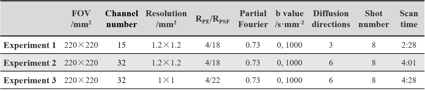
Data acquisitionWith institutional review board approval, data were obtained from 3 healthy volunteers on a 3T Philips Achieva scanner (Philips Healthcare, Best, The Netherlands) using the PSF-EPI sequence. Three experiments were carried out to test the performance of the proposed method. The first experiment used a 15-channel head coil to validate the proposed method. The second and the third experiments used a 32-channel head coil to test the method with different resolution data and DTI quantification. The scan parameters are shown in Table 1.
Image reconstruction
Images were reconstructed using the Tilted-CAIPI method and the proposed method, respectively. Sensitivity maps were calculated from b0 data using ESPIRIT8 and colored FA maps were calculated using FSL9.
Results and discussion
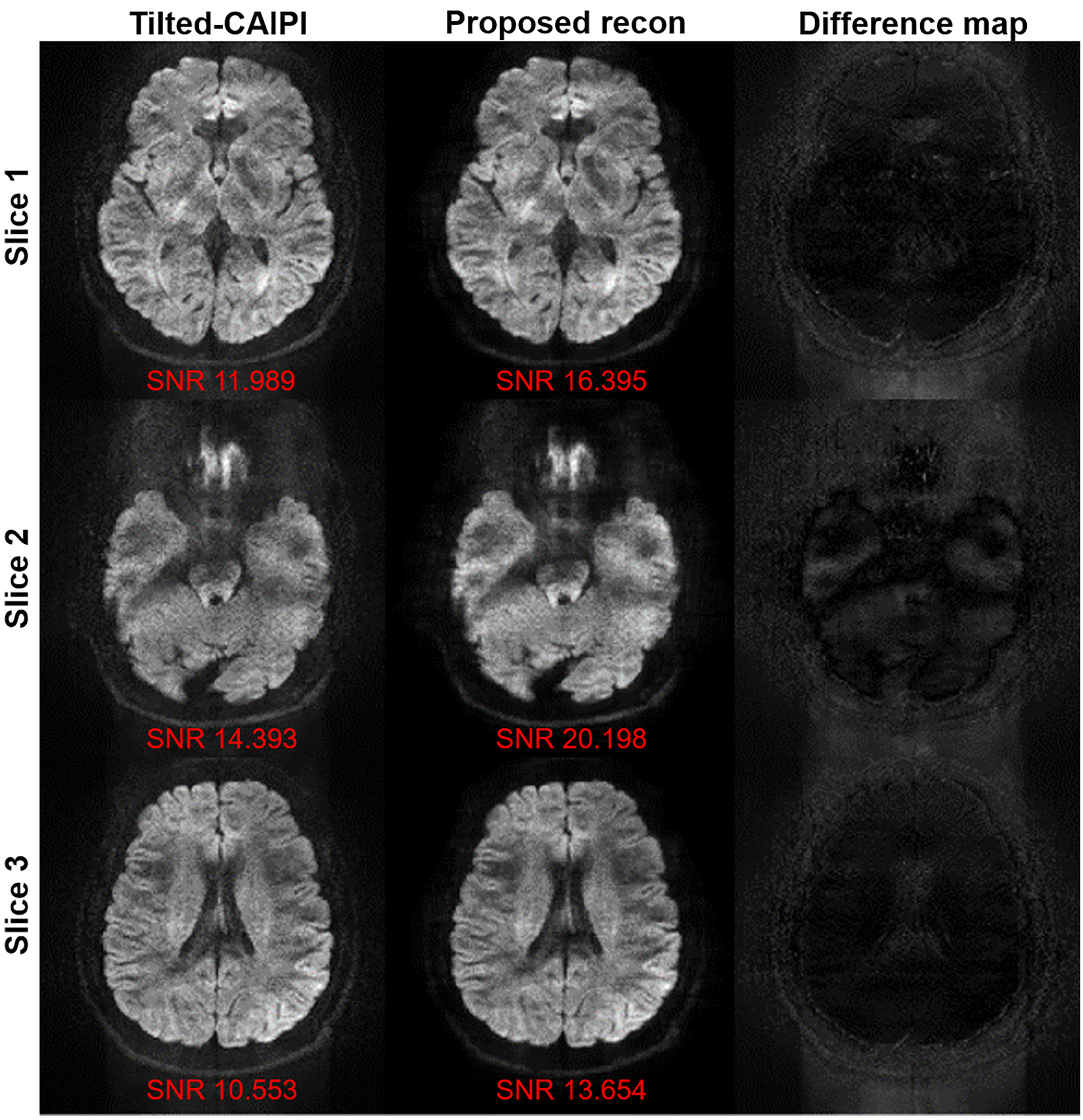
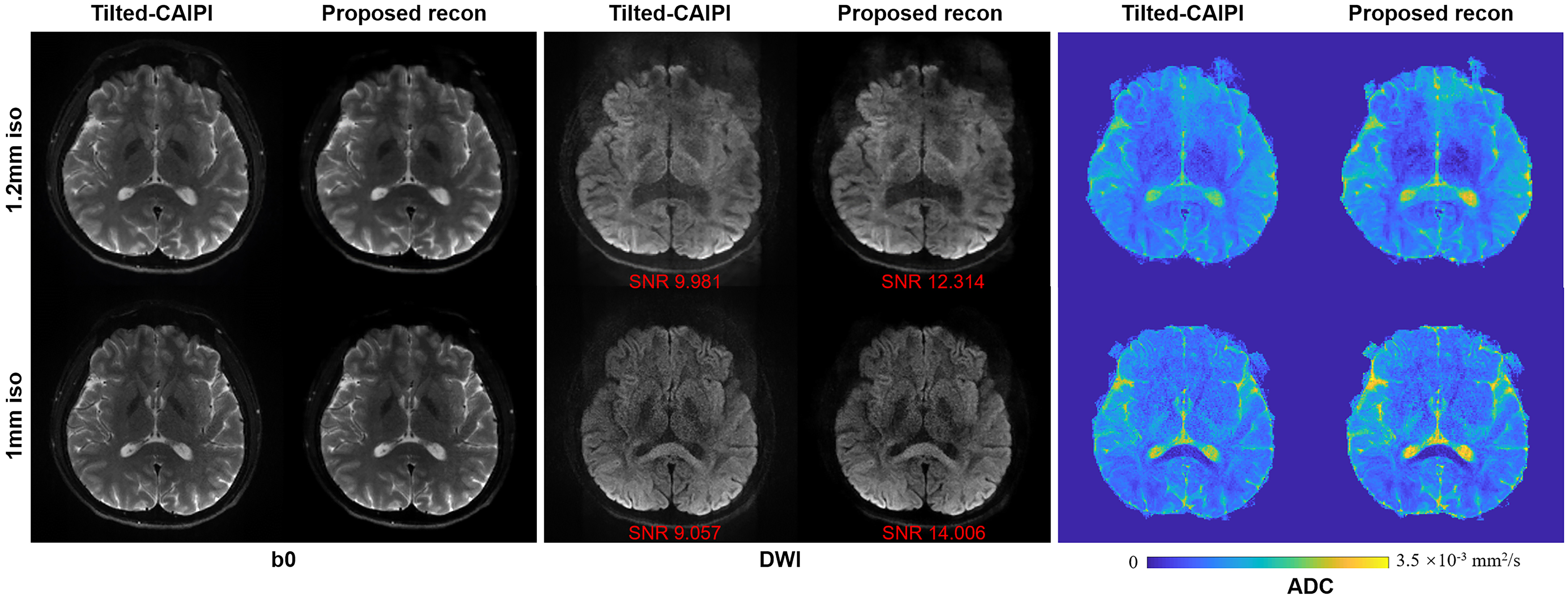
Figure 2 shows 3 slices of the DWI images from experiment 1. Compared to using Tilted-CAIPI, the noise levels of reconstructed images using the proposed method decrease and the SNRs improve since the low rank constraint jointly updates all 2D images along the ky dimension.Similar improvements can be found when the image resolution increases. Figure 3 shows the reconstructed images from experiments 2 and 3. The 1 mm isotropic resolution DWI image reconstructed from Tilted-CAIPI (middle panel) is noisier than that from the proposed recon. The SNR values also indicate the improvement for both resolution DWI images when using the proposed reconstruction method. ADC values of the brain tissues lie within the normal range of healthy adults.
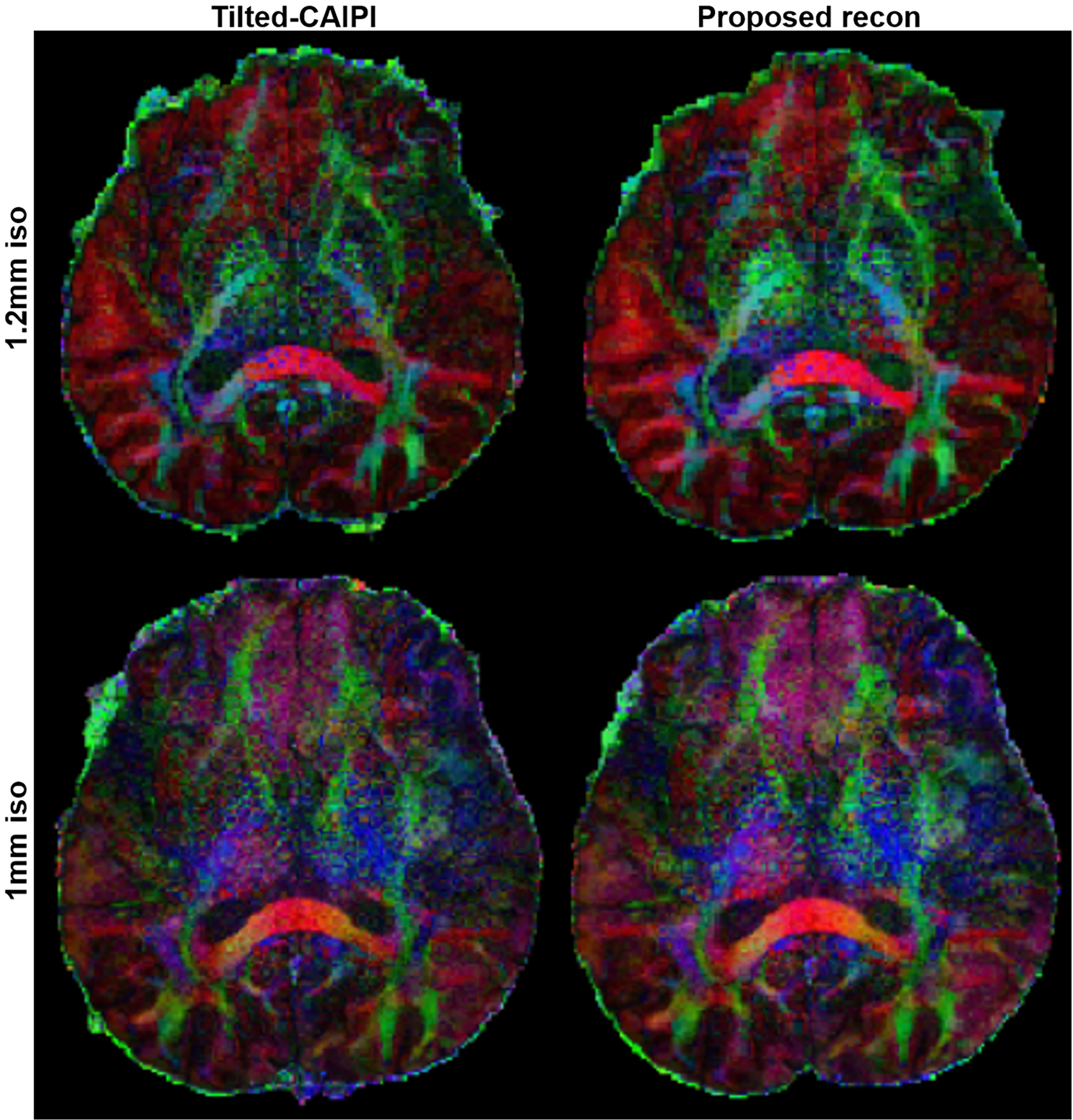
We further examine the proposed method in DTI. The colored FA maps are shown in Figure 4. Compared to using Tilted-CAIPI, the obtained cFA maps using the proposed method are visually better, as the noise levels decrease. Additionally, the small structures can also be preserved.
Conclusion
The proposed low-rank based reconstruction algorithm can improve the DW PSF-EPI images obtained from Tilted-CAIPI with increased SNR. It may have potentials to improve the reliability of Tilted-CAIPI accelerated PSF-EPI.Acknowledgements
No acknowledgement found.References
1. Bammer R. Basic principles of diffusion-weighted imaging. Eur J Radiol 2003;45(3):169-184.
2. Skare S, Newbould RD, Clayton DB, Bammer R. Propeller EPI in the other direction. Magn Reson Med 2006;55(6):1298-1307.
3. Chen NK, Guidon A, Chang HC, Song AW. A robust multi-shot scan strategy for high-resolution diffusion weighted MRI enabled by multiplexed sensitivity-encoding (MUSE). Neuroimage 2013;72:41-47.
4. Mani M, Jacob M, Kelley D, Magnotta V. Multi-shot sensitivity-encoded diffusion data recovery using structured low-rank matrix completion (MUSSELS). Magnetic Resonance in Medicine 2017;78(2):494-507.
5. In MH, Speck O. Highly accelerated PSF-mapping for EPI distortion correction with improved fidelity. Magn Reson Mater Phy 2012;25(3):183-192.
6. Dong ZJ, Wang FYX, Reese TG, et al. Tilted-CAIPI for highly accelerated distortion-free EPI with point spread function (PSF) encoding. Magnetic Resonance in Medicine 2019;81(1):377-392.
7. Porter DA, Heidemann RM. High Resolution Diffusion-Weighted Imaging Using Readout-Segmented Echo-Planar Imaging, Parallel Imaging and a Two-Dimensional Navigator-Based Reacquisition. Magnetic Resonance in Medicine 2009;62(2):468-475.
8. Uecker M, Lai P, Murphy MJ, et al. ESPIRiT-An Eigenvalue Approach to Autocalibrating Parallel MRI: Where SENSE Meets GRAPPA. Magnetic Resonance in Medicine 2014;71(3):990-1001.
9. Jenkinson M, Beckmann CF, Behrens TE, Woolrich MW, Smith SM. Fsl. Neuroimage 2012;62(2):782-790.
Figures