4340
Diffusion Weighted Imaging using PROPELLER Acquisition and a Deep Learning based Reconstruction1Global MR Applications & Workflow, GE Healthcare, Houston, TX, United States, 2Global MR Applications & Workflow, GE Healthcare, New York, NY, United States, 3Global MR Applications & Workflow, GE Healthcare, Waukesha, WI, United States, 4Global MR Applications & Workflow, GE Healthcare, Calgary, AB, Canada, 5Global MR Applications & Workflow, GE Healthcare, Tucson, AZ, United States, 6Global MR Applications & Workflow, GE Healthcare, Boston, MA, United States, 7Global MR Applications & Workflow, GE Healthcare, Menlo Park, CA, United States
Synopsis
PROPELLER DWI, a FSE based DWI method, is increasingly used to reduce susceptibility artifacts and motion artifacts. Multi-shot Echo-Planar diffusion method also can reduce susceptibility artifacts, but PROPELLER DWI shows better image quality where susceptibility artifacts are most problematic, such as in skull base, head-neck and pelvis. However, the acquisition time is often longer compared to ms-DW-EPI, therefore SNR is usually compromised to reduce acquisition time. In this work, we evaluated a deep-learning based reconstruction method (DL Recon PROP) intended to improve image quality and ADC measurements by reducing the noise and artifacts without increasing acquisition time.
Introduction
PROPELLER DWI (1,2), a Fast-Spin-Echo (FSE-) based diffusion weighted imaging (DWI) is increasingly used to reduce susceptibility artifacts and motion artifacts. The in-plane resolution is also improved compared to single-shot DW methods. Multi-shot Echo-Planar diffusion method (ms-DW-EPI) (3) also can reduce susceptibility artifacts, but PROPELLER DWI shows better image quality where susceptibility artifacts are most problematic, such as in skull base, head-neck and pelvis with metal implant (4). However, the acquisition time is often longer compared to ms-DW-EPI, therefore SNR is usually compromised to reduce acquisition time. The purpose of this work was to evaluate a convolutional neural network (CNN) based reconstruction method to improve the PROLLER DW image quality and ADC measurements by reducing the noise and artifacts without increasing acquisition time.Methods
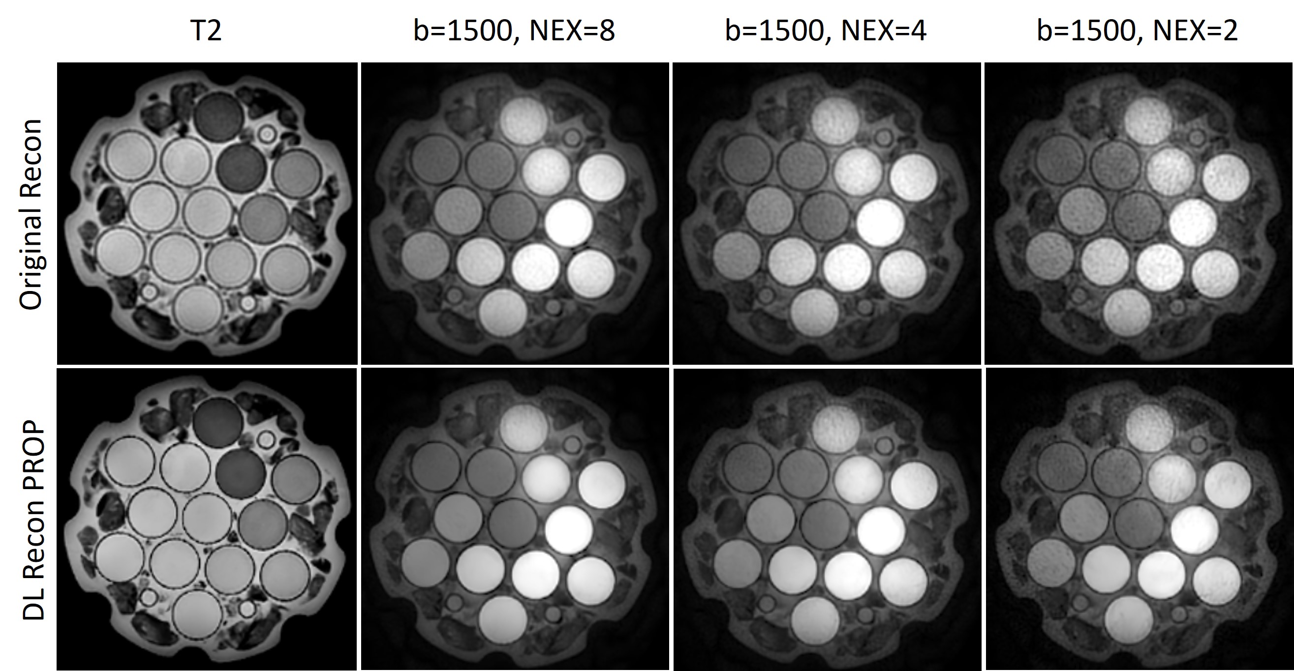
The CNN based deep-learning network was trained for PROPELLER reconstruction ((DL Recon PROP)) with over 10,000 high SNR and high spatial resolution images. The DL Recon PROP was designed to improve the SNR and spatial resolution, and was embedded into the conventional reconstruction pathway to generate two sets of image series from a single set of raw MR data. The proposed DL Recon PROP method was first evaluated with a standard NIST diffusion phantom to demonstrate the artifacts and noise removal as well as quantifying the ADC measurements. The diffusion phantom images were acquired using PROPELLER DWI with NSA = 8, 4 and 2, and a parallel acceleration factor of 2. Since diffusion phantoms have long T2 relaxation times compared to most soft tissues, the receiver bandwidth was intentionally increased to reduce SNR for evaluation. Finally, the proposed DL Recon PROP method was evaluated and compared against conventional PROPELLER DWI and ms-DW-EPI in the brain, prostate and female pelvis of 5 healthy volunteers with IRB approval and written informed consent. All images were acquired at a 3T GE MR scanner (GE Healthcare, Waukesha, WI).Results
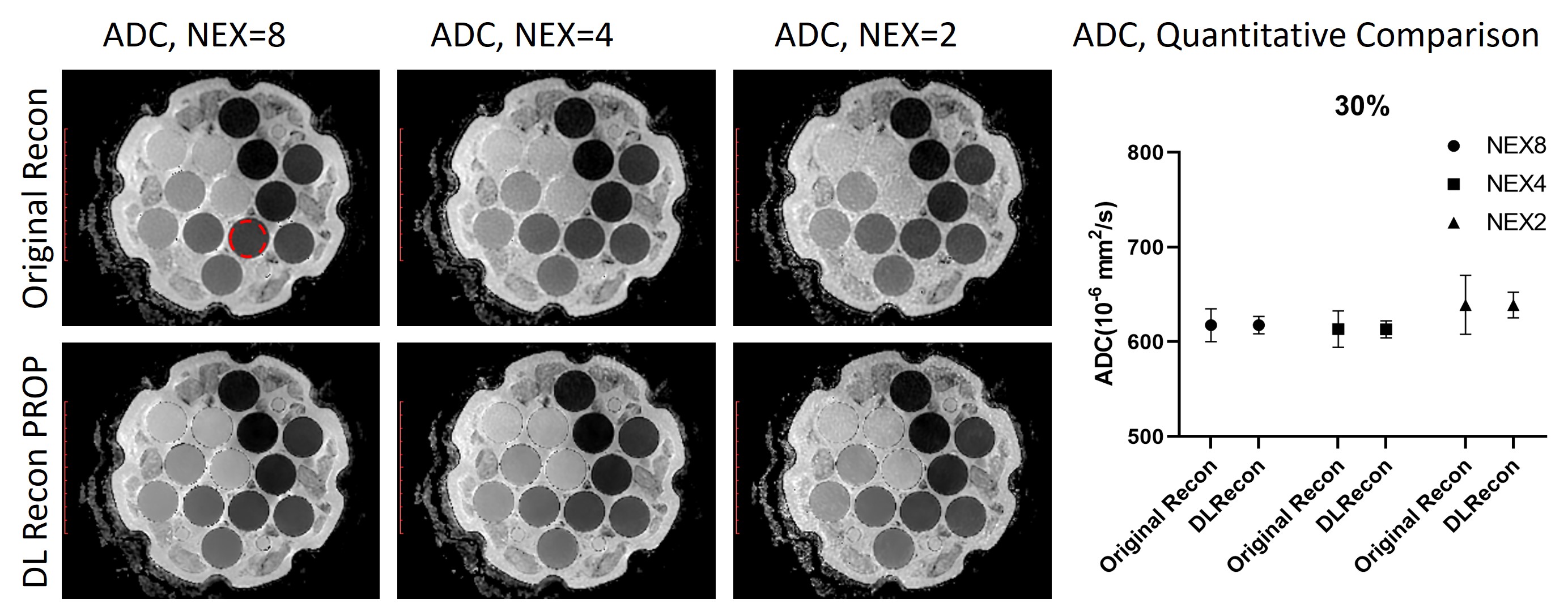
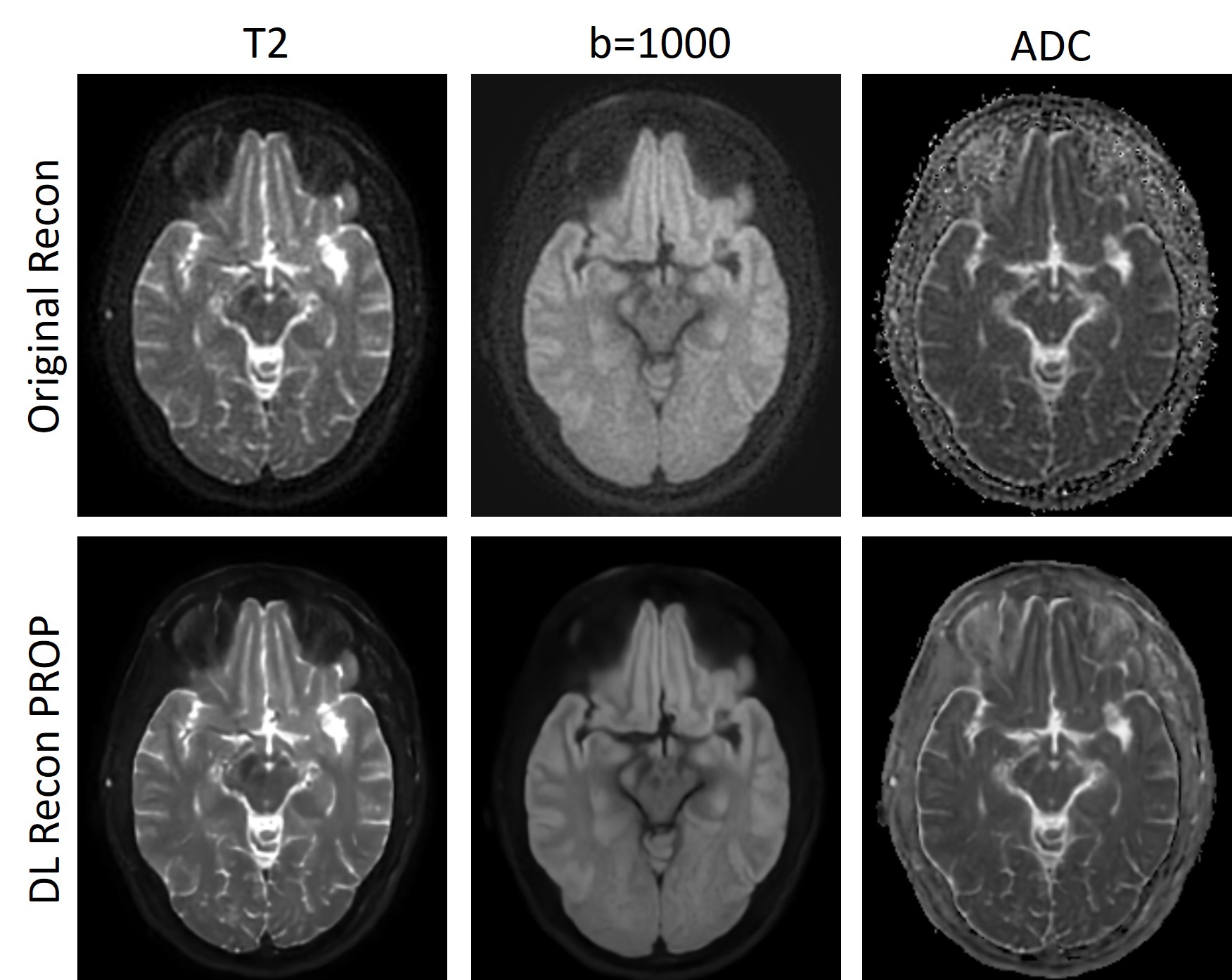
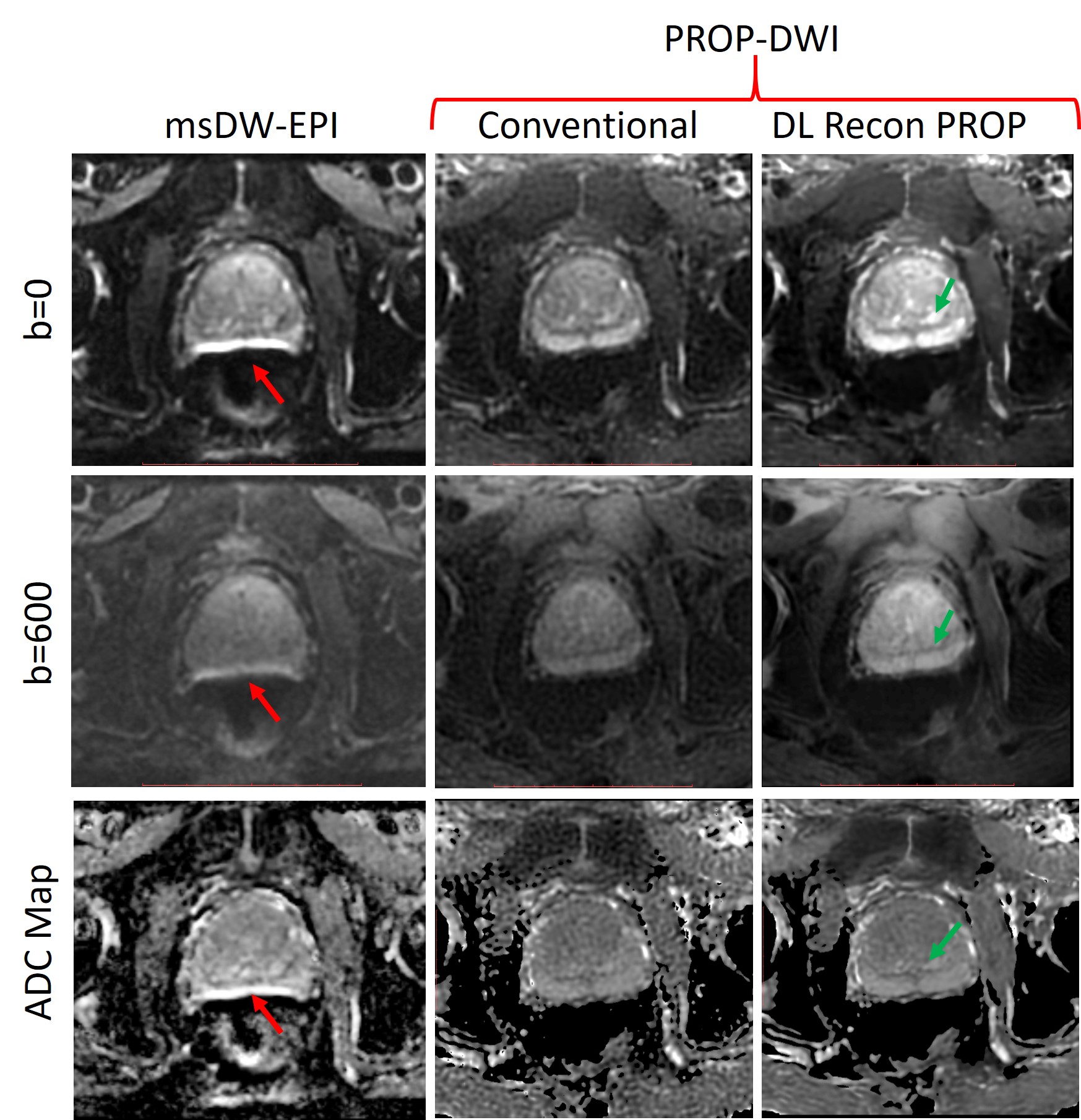
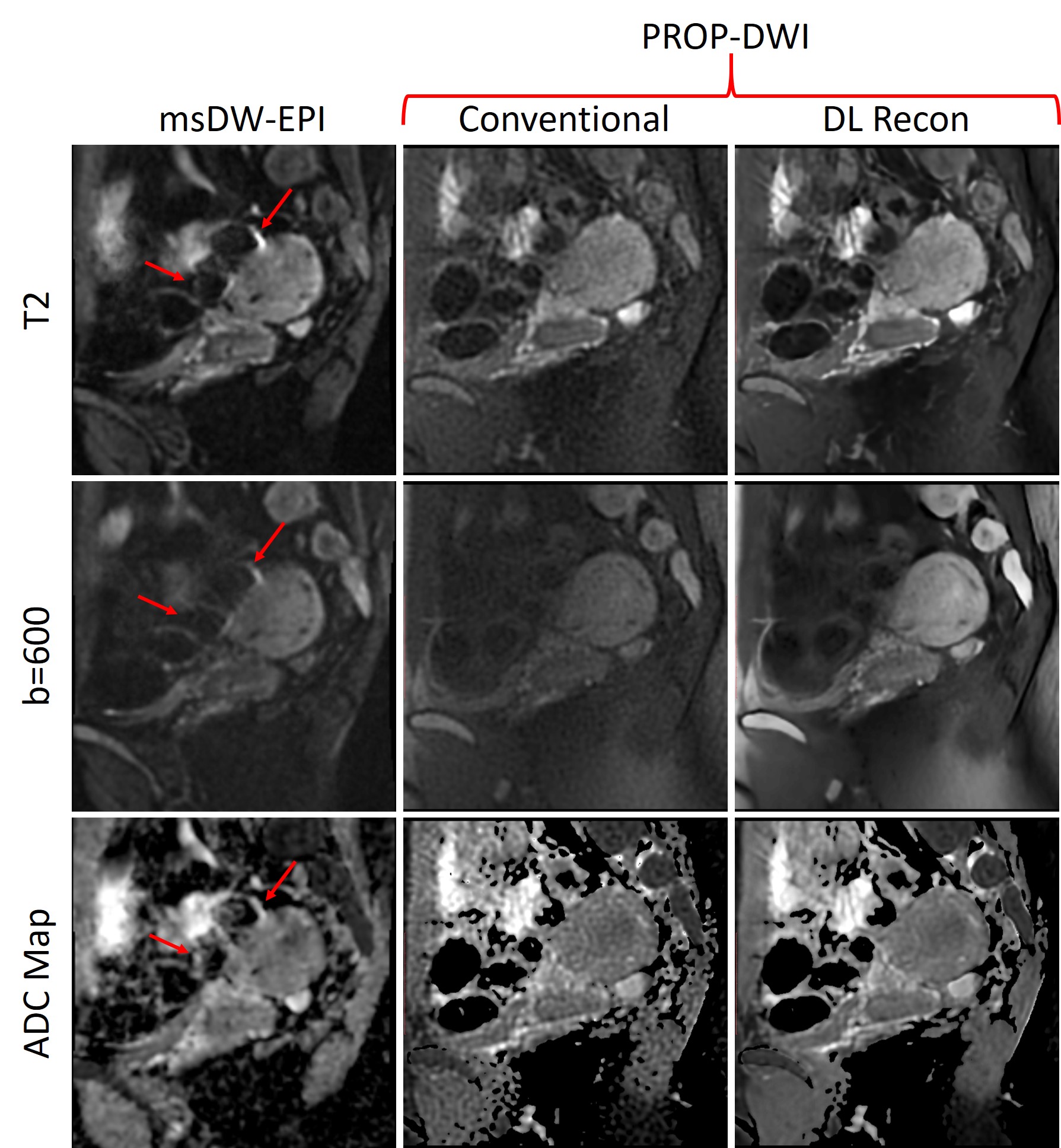
The diffusion phantom images reconstructed with conventional reconstruction method and DL Recon PROP method were shown in Figure 1. Compared to the conventional reconstruction method, DL Recon PROP increased the SNR and image sharpness, as shown in Fig.1 (T2 image). As expected, the SNR of b1500 images was reduced by reducing NEX factor. However, the apparent SNR was increased using DL Recon PROP, and the b1500 image with a NEX of 2 and DL Recon PROP was not visually inferior to b1500 image with a NEX of 8 and conventional reconstruction method. DL Recon PROP also improved ADC measurements, as shown in Figure 2. ADC maps were generated from Figure 1 with both conventional reconstruction method and DL Recon PROP. The ADC maps reconstructed with DL Recon PROP also showed improved SNR and image sharpness. Compared to the conventional reconstruction method, DL Recon PROP did not change the mean ADC values across different NEX or different concentrations of PVP, as shown in the representative plot of the measured ADC values from vial with 30% PVP. DL Recon PROP produced the same mean ADC values as the conventional reconstruction method, but with comparatively smaller standard deviation. The representative PROPELLER T2 and b1000 brain images of a healthy volunteer were shown in Figure 3. DL Recon PROP reduced noise and improved sharpness of both T2 and b1000 images, resulting in a better ADC estimates with smaller variations and fewer artifacts compared to the conventional reconstruction method. Multishot DW-EPI and PROPELLER DWI prostate (Figure 4) and pelvis (Figure 5) images of healthy volunteers were acquired without endorectal coil and gel. Multishot DW-EPI images were acquired with 3 shots and reconstructed with original reconstruction methods. PROPELLER T2 and high b-value images were reconstructed using both conventional reconstruction method and DL Recon PROP method. The B0 inhomogeneities in the anatomies were large due to the presence of multiple air-tissue interfaces, resulting in large susceptibility artifacts. Susceptibility artifacts (Figure 4 and 5, red arrows) were shown in DW-EPI images, but not in PROPELLER images. The PROPELLER images reconstructed using conventional method showed low SNR. Howerver, DL Recon PROP significantly improved SNR and image sharpness of both T2 and high b-value images, resulting in a better ADC estimates with smaller variations. The peripheral zone (Figure 4, red arrow) is more clearly visualized in the DL Recon PROP images than in the conventional PROPELLER DWI images.Conclusion and Discussion
In this work, we demonstrated that DL Recon PROP method to improve the image quality of DW PROPELLER images without increasing acquisition time. In addition to increasing apparent SNR, DL Recon PROP also improved image sharpness. DL Recon PROP will not change the mean ADC values, while make it more precise.Acknowledgements
No acknowledgement found.References
1. James G. Pipe, Nicholas Zwart. Turboprop: Improved PROPELLER Imaging. MRM. 2006;55(2):380-385
2. Xiaoli Zhao, Zhiqiang Li, Ajeetkumar Gaddipati. PROPELLER DUO: Applied to Diffusion-Weighted Imaging. ISMRM, 2009
3. Chen, N.-K., Guidon, A., Chang, H.-C., Song, A.W. A robust multi-shot scan strategy for high-resolution diffusion weighted MRI enabled by multiplexed sensitivity-encoding (MUSE). NeuroImage. 2013;72:41-47
4. Czarniecki M, Caglic I, Grist JT, Gill AB, Lorenc K, Slough RA, Priest AN, Barrett T. Role of PROPELLER-DWI of the prostate in reducing distortion and artefact from total hip replacement metalwork. Eur J Radiol. 2018;102:213-219
Figures