4339
SENSE-based Multi-shot DWI Reconstruction with Extra-navigated Rigid Motion and Contrast Correction for Brain EPI1Institute for Signal Processing, University of Luebeck, Luebeck, Germany, 2Philips Research Europe, Hamburg, Germany, 3Department of Radiology, LUMC, Leiden, Netherlands
Synopsis
We propose an extra-navigated SENSE-based multi-shot DWI reconstruction algorithm that comprises navigator-based phase and rigid in-plane motion corrections at fast reconstruction times. Furthermore, this approach exploits the low-resolution navigator signal to perform diffusion contrast corrections explicitly within the model. The extra-navigated method is compared in-vivo to a self-navigated reference algorithm. The extra-navigated motion estimation from low-resolution navigator data yields decent reconstructions which perfectly coincide with self-navigated results. Moreover, extra-navigation allows for fast reconstruction at the cost of lower scan efficiency and appears to be more robust for strong motion corruption and high segmentations.
Introduction
Multi-shot DWI techniques offer promising potential to improve current state-of-the-art diffusion-weighted imaging (DWI) and diffusion tensor imaging (DTI) applications1 in terms of SNR and spatial resolution. However, multi-shot DWI is prone to shot-specific variations from tiny physiological motion2 as well as inter-shot gross motion. Supported by parallel imaging techniques3,4, self-navigated5,6 and navigator-free7 reconstruction algorithms have been proposed to overcome the shot-specific variations. Besides those data-driven approaches, extra-navigated methods provide robust alternatives at the cost of decreased scan efficiency, sampling a 2D low-resolution navigator image after a second refocusing pulse8. Navigator-based multi-shot DWI reconstructions effectively capture shot phase variations8 and rigid in-plane motion9 including contrast corrections to account for the changed effective diffusion direction after rotational motion. This work presents a fast extra-navigated multi-shot DWI reconstruction algorithm based on SENSE and further exploits the navigator signal for low-resolution diffusion contrast corrections.Theory
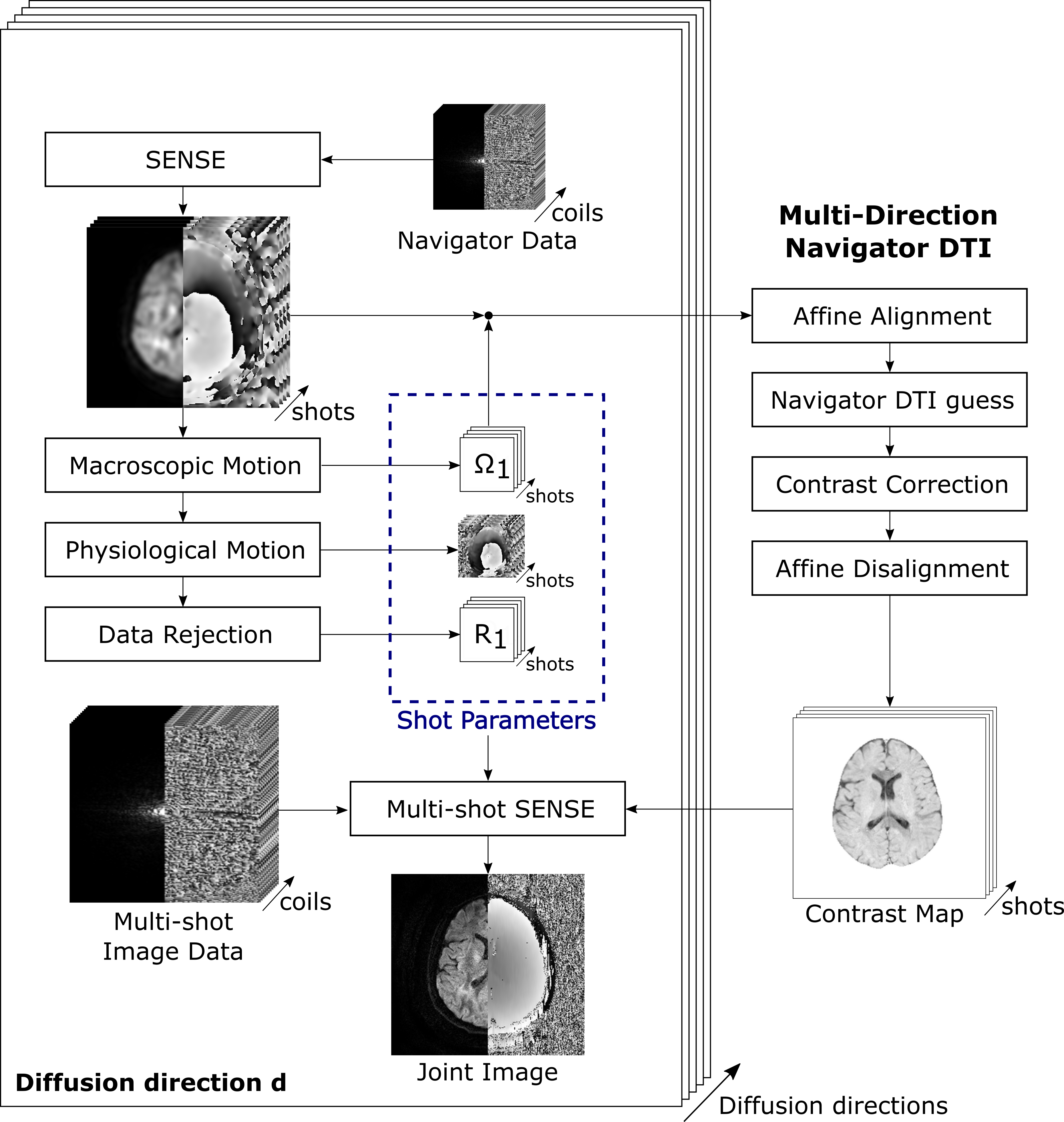
The extra-navigated macroscopic motion-corrected algorithm solves a multi-shot DWI problem that differs from a previous approach9 by introducing navigator-based contrast corrections into the model and adopting a SENSE4-based formulation:$$ \hat{\mathbf{\rho}} = \underset{\mathbf{\rho}}{\mathrm{argmin}} \quad \frac{1}{2} \sum_{i \in I} \lVert F_i S \Omega_i \Phi_i C_i \; \mathbf{\rho} - \mathbf{d}_i \rVert_2^2 + \alpha \lVert \mathbf{\rho} \rVert_2^2,$$
where $$$F_i$$$, $$$\Omega_i$$$, $$$\Phi_i$$$ and $$$C_i$$$ are the Fourier, macroscopic motion, physiological motion and contrast correction operator of shot $$$i$$$, respectively. $$$\mathbf{\rho}$$$ is the joint image, $$$S$$$ the sensitivity operator, $$$\mathbf{d}_i$$$ the i-th shot data, $$$\alpha$$$ the Tikhonov regularization parameter and $$$I$$$ the set of included shots from $$$N_i$$$ total shots to allow for data rejection. This model is solved for every diffusion direction $$$k$$$ of $$$N_k$$$ total directions.
Algorithms
First, the navigator data is upsampled to full resolution by a triangular window and reconstructed using SENSE4. Next, the rigid motion, shot phases and data rejection criteria are subsequently calculated from the navigator images. Then, diffusion tensor estimates are calculated from the navigator magnitudes after affine in-plane alignment to calculate shot-wise low-resolution diffusion contrast correction maps according to the estimated rotation. Note that this DTI analysis is performed using all $$$N_k N_i$$$ navigator shots with their respective rotation-corrected diffusion direction6,9. As there have not been any shot combinations up to this DTI estimation, unlike the approach by Dong et al9, the proposed approach remains unbiased. Finally, the multi-shot model is solved for the joint image estimate $$$\hat{\mathbf{\rho}}$$$ using conjugate gradients (CG). The algorithm is visualized in Fig. 1.This algorithm is evaluated with respect to IRIS8 and a self-navigated algorithm10. IRIS is an extra-navigated approach that only includes phase operators $$$\Phi_i$$$ into the multi-shot model, whereas the iterative self-navigated approach also corrects for rigid motion $$$\Omega_i$$$. Partial Fourier is included by homodyne reconstruction. Image registration is performed using a normalized gradient field metric11. The shot with the highest correlation to all other shots is chosen as the reference shot for registration and data rejection. Shots below 95% correlation are excluded.
Methods
Multi-shot echo-planar diffusion data was acquired with an extra 2D navigator using a twice-refocused Stejskal-Tanner sequence8. The data was obtained from 8 healthy volunteers on a 3T Philips Ingenia using a 13-channel head coil. Informed consent was obtained according to the rules of the institution. DTI data was acquired with {4, 5} shots, b = {0, 1000} s/mm2 in 15 diffusion directions with a resolution of 1x1x4 mm3. The DTI experiments were performed twice, first the subjects remained still and second continuous in-plane motion was performed. Fractional anisotropy (FA) maps were calculated using Dipy12 after affine alignment.Results
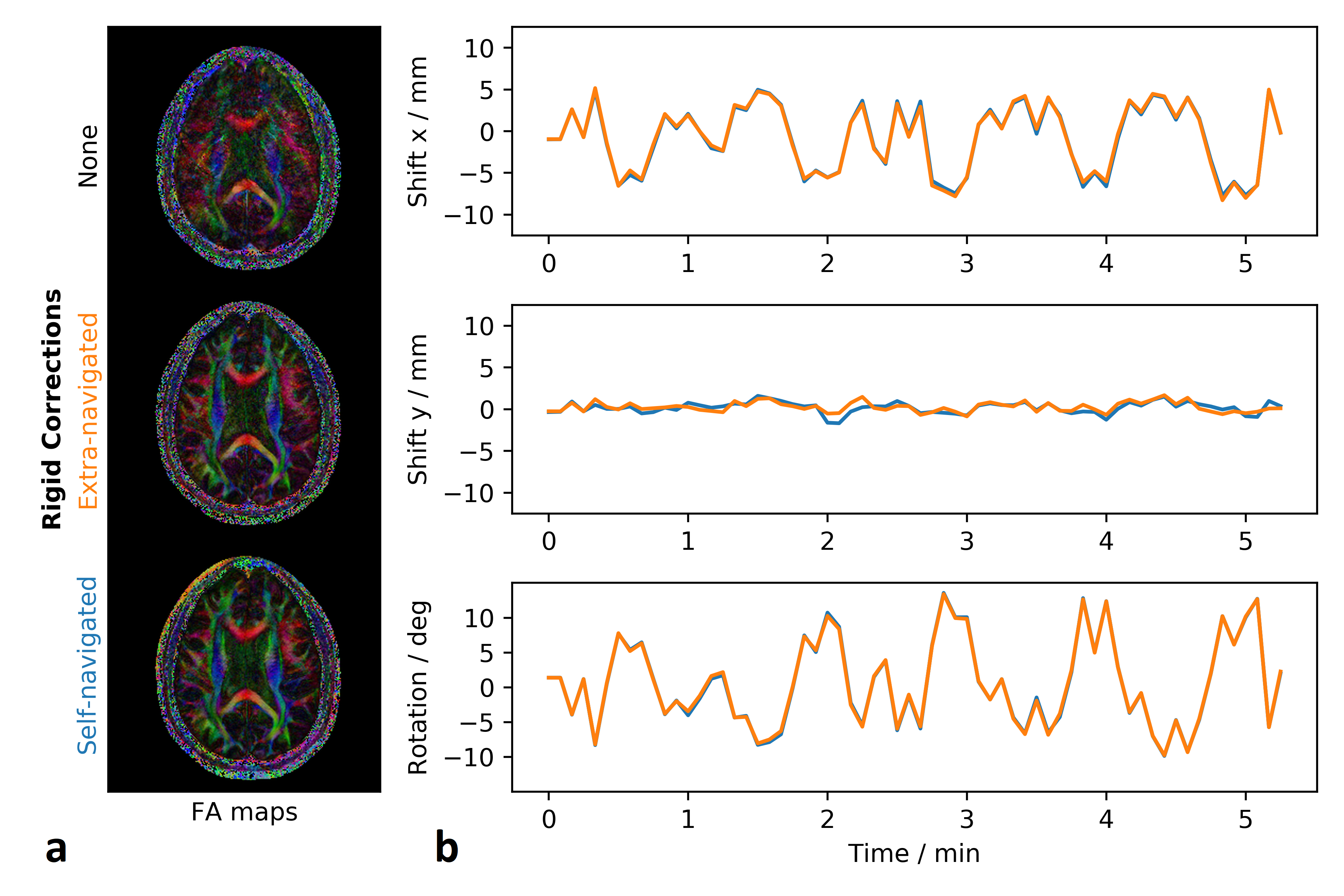
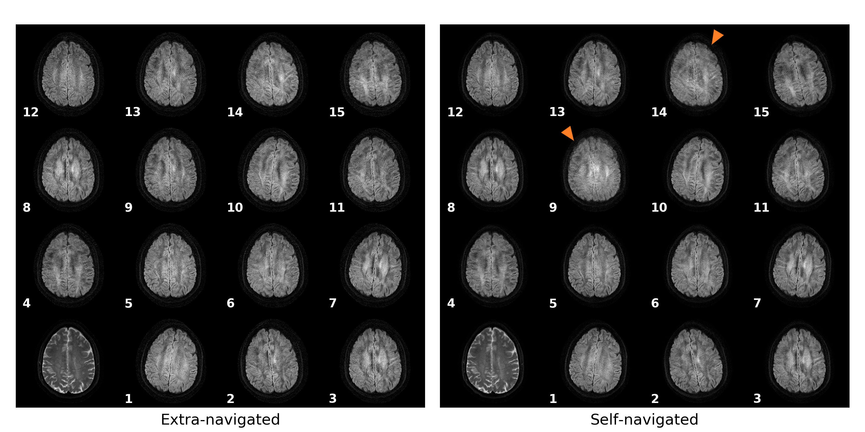
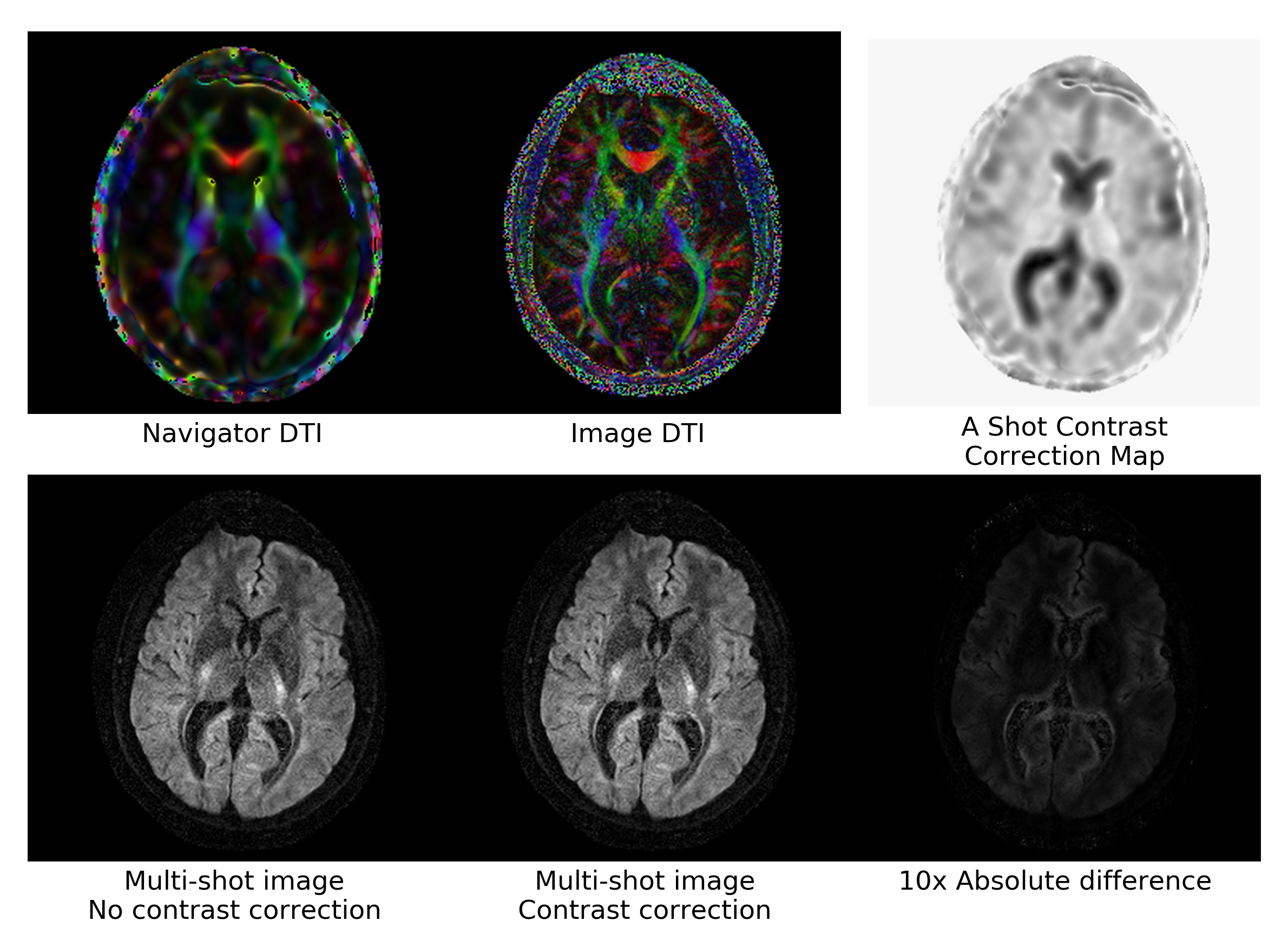
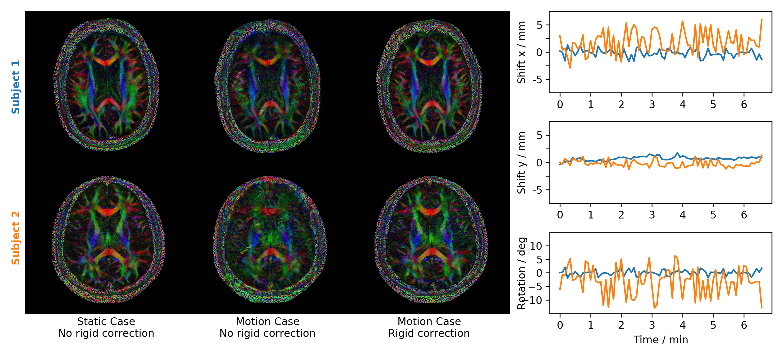
Figure 2 compares the proposed extra-navigated to the self-navigated10 approach for a motion-corrupted dataset. Figure 3 shows the full sets of 5-shot DWI reconstructions within a DTI dataset for a severely motion-corrupted case. The data was registered to the mean rigid motion direction within each multi-shot group. The multi-shot images thus remain misaligned. A diffusion contrast example is provided in Fig. 4. Figure 5 compares extra-navigated DTI reconstructions under static and dynamic conditions. The extra-navigated method took on average about 15 s and 17 s with contrast correction disabled and enabled, whereas the self-navigated approach needed on average 52 s with 55 iterations.Discussion
The rigid motion estimates of the self- and the extra-navigated approaches coincide remarkably well (Fig. 2). The low-resolution navigators prove to enable sufficiently accurate macroscopic motion estimation. In extreme cases with severe rigid motion and high segmentation, the extra-navigated approach was found to be more robust due to the reliable low-resolution information and the equality of the navigator trajectories for each shot (Fig. 3). The contrast corrections are in a range around and below 5%. The evaluation of the contrast correction in simulations is subject to future research (Fig. 4). Both approaches could be further extended by subsequent high-resolution diffusion contrast corrections, as previously suggested9. The extra-navigated approach recovers important structural brain information in the presence of strong in-plane motion and provides fast reconstructions and robust navigators at the cost of less scan efficiency (Fig. 5).Conclusion
The proposed SENSE-based extra-navigated multi-shot approach efficiently recovers decent diffusion images and proves to be a robust alternative to self-navigated algorithms, especially for strong in-plane motion and high segmentations paving the way for future clinical use.Acknowledgements
No acknowledgement found.References
1. Wu W and Miller KL. Image formation in diffusion MRI: A review of recent technical developments: Review of Image Formation in dMRI. JMRI. 2017;46(3):646–662.
2. Miller KL and Pauly JM. Nonlinear Phase Correction for Navigated Diffusion Imaging. MRM. 2003;50:343-353.
3. Roemer PB, Edelstein WA, Hayes CE, Souza SP, Mueller OM. The NMR phased array. MRM. 1990;16(2):192–225.
4. Pruessmann et al. SENSE: sensitivity encoding for fast MRI. MRM. vol. 1999;42(5):952–962.
5. Guo et al. POCS‐enhanced inherent correction of motion‐induced phase errors (POCS‐ICE) for high‐resolution multishot diffusion MRI. MRM. 2016;75(1):169-180.
6. Guhaniyogi et al. Motion Immune Diffusion Imaging Using Augmented MUSE for High-Resolution Multi-Shot EPI. MRM. 2016;75:639-652.
7. Mani M, Jacob M, Kelley D, Magnotta V. Multi-shot sensitivity-encoded diffusion data recovery using structured low-rank matrix completion (MUSSELS): Annihilating Filter K-Space Formulation for Multi-Shot DWI Recovery. MRM. 2017;78(2):494-507.
8. Jeong H-K, Gore JC, Anderson AW. High-resolution human diffusion tensor imaging using 2-D navigated multishot SENSE EPI at 7 T. MRM. 2013;69(3):793-802.
9. Dong Z, Wang F, Ma X, Dai E, Zhang Z, Guo H. Motion-corrected k-space reconstruction for interleaved EPI diffusion imaging: Motion Correction for iEPI DWI. MRM. 2018;79(4):1992-2002.
10. Steinhoff M, Nehrke K, Mertins A, Börnert P. Multi-shot Diffusion EPI Reconstruction with Iterative Rigid Motion-correction and Motion-induced Phase-correction for Brain Imaging. In: Proceedings of the 27th Joint Annual Meeting of ISMRM. Montréal, QC, Canada; 2019.
11. Kabus S and Lorenz C. Fast elastic image registration. Medical Image Analysis for the Clinic: A Grand Challenge. 2010:81-89.
12. Garyfallidis E, Brett M, Amirbekian B, et al. Dipy, a library for the analysis of diffusion MRI data. Frontiers in neuroinformatics. 2014;8:8.
Figures