4337
Readout-Segment Echo-Planar Imaging of Prostate, a Strategy to Reduce Geometrical Distortion in Prostate Diffusion Weighted Imaging1Abdominal Radiology, David Geffen School of Medicine at UCLA, Los Angeles, CA, United States
Synopsis
Single-Shot Echo-Planar Imaging (ssEPI)is highly susceptible to T2* blurring and geometrical distortion. This study aimed to compare the image quality between ssEPI and Readout-Segment Echo-Planar Imaging (rsEPI) for acquiring prostate DWI on 162 patients. Geometrical distortion was ranked on ssEPI and rsEPI using a five-point scale. The geometrical distortion was significantly less observed in rsEPI compared to ssEPI (P<0.01). Geometrical distortion scores of three and higher were observed in 30 individuals in ssEPI, with all having scores < 3 on rsEPI. In conclusion, using rsEPI for DWI acquisition may augment or replace ssEPI on 3T prostate mpMRI.
Introduction
Diffusion-Weighted Imaging (DWI) is a key component of risk assessment in multiparametric MRI (mpMRI) for prostate cancer (Pca) detection. Single-Shot Echo-Planar Imaging (ssEPI)is considered as the method of choice for acquisition of prostate DWI. This technique is highly susceptible to T2* blurring and geometrical distortion, notably at stronger (e.g. 3 Tesla) magnetic fields. This study aimed to compare the image quality between ssEPI and Readout-Segment Echo-Planar Imaging (rsEPI) for acquiring prostate DWI. We aimed to examine whether rsEPI can lower geometrical distortion in prostate DWI.Methods
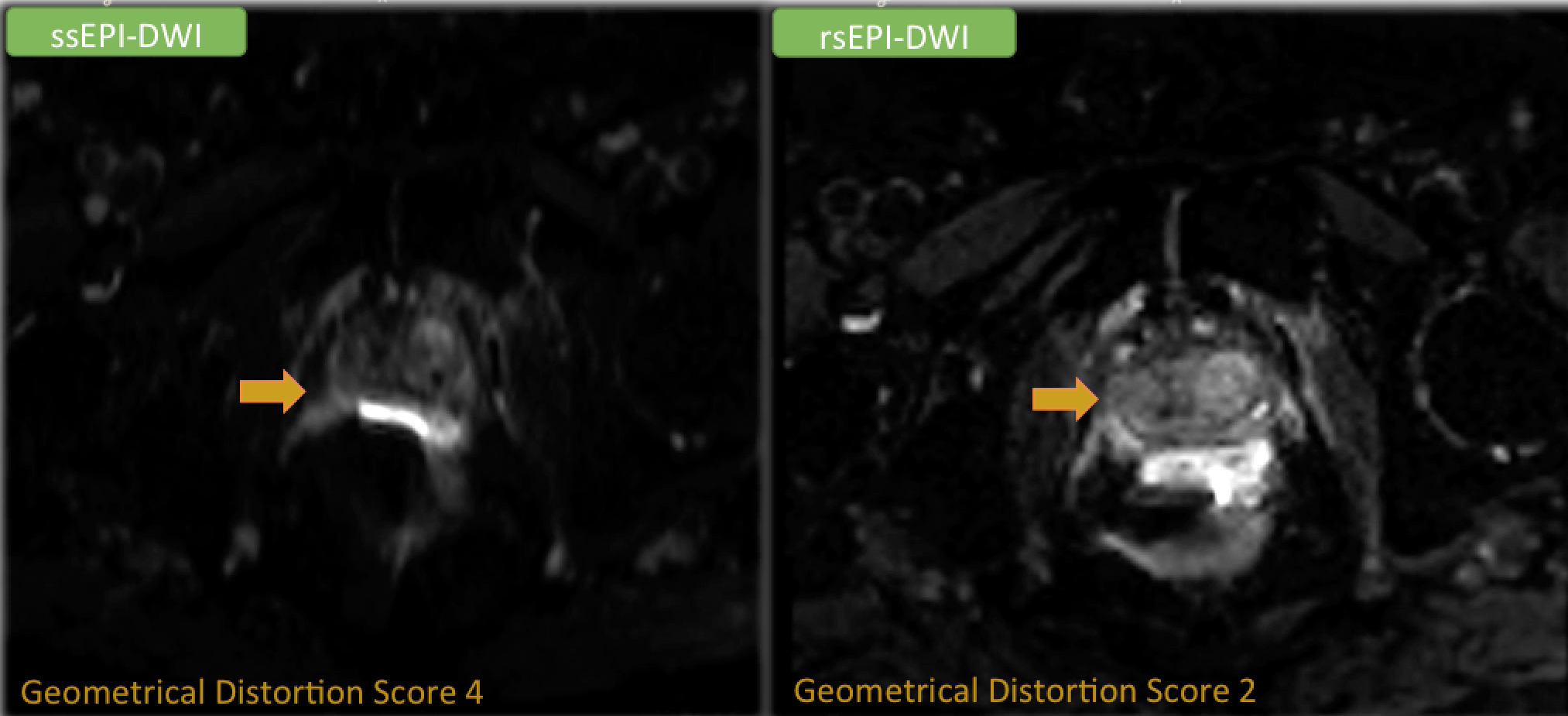
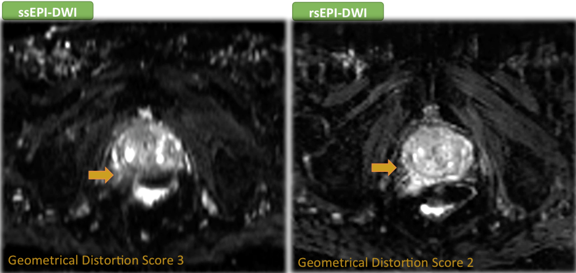
This IRB-approved, HIPAA-compliant, single-center study included 162 consecutive patients who underwent 3Tesla prostate multiparametric MRI (mpMRI) between January and April 2019. Patients with history of prior pelvic surgery or radiotherapy were excluded. Prostate MRI was acquired using a 3 Tesla MR scanner with a pelvic external phased-array coil. All MRIs followed the same standard protocol, which included two-dimensional turbo spin-echo T2-weighted imaging, dynamic contrast-enhanced (DCE) imaging and DWI using both ssEPI and rsEPI. The following image parameters were used for acquisition of ssEPI and rsEPI, respectively: b values: 0, 100, 400, 800 and calculated high b value of 1400 s/mm2vs. 50 and 800 s/mm2, TR/TE: 4800/80 vs. 4530/57 milliseconds, in-plane resolution: 1.6 vs. 2 mm2, field of view: 260 vs. 220 mm and matrix size/number of segments: 160*94/ 1 vs. 110*110/ 5. Three radiologists, blinded to clinical data, independently reviewed each MRI to rank geometrical distortion on ssEPI and rsEPI using a five-point scale as followed: 1:no perceptible artifact, 2:minimal susceptibility artifact (SA), 3:mild SA, obscuring <50% of peripheral zone (PZ), 4:moderate SA, obscuring >50% of PZ, and 5:severe SA, affecting peripheral and transition zones. The degree of rectal gas was also ranked on DCE images using a five-point scale. SPSSv.16 was used to run Wilcoxon signed rank test for comparison of rankings and weighted Kappa test to assess inter-rater agreement.Results
The inter-reader agreement for scoring image distortion in ssEPI and rsEPI were 0.85 and 0.79, respectively. Mean image distortion scores were 1.61 and 1.16 in ssEPI and rsEPI techniques, respectively. The geometrical distortion was significantly less observed in rsEPI compared to ssEPI, regardless of the rectal gas degree (P<0.01) and when stratified by rectal gas scoring (P<0.05). Geometrical distortion scores of three and higher were observed in 30 individuals in ssEPI, with all having scores < 3 on rsEPI.Discussion
A number of studies have found using rsEPI to decrease geometrical distortion in diffusion-weighted imaging of several parts of body.1-3 Our data showed outperformance of rsEPI compared to ssEPI for reducing image distortion in prostate diffusion-weighted imaging.Single shot EPI requires long echo train and long TE to encode full k space within 1 echo signal intensity, therefore it is susceptible to T2* blurring and signal drop out at air-tissue interface. Furthermore, due to shorter T2* relaxation time and increased matrix size, the sensitivity to susceptibility artifact doubles at 3 Tesla compared to 1.5 T MR scanner. rsEPI, in comparison, uses a higher bandwidth per pixel with shorter effective TE, leading to reduced T2 shine-through effect.Conclusion
rsEPI performed significantly better in producing DWI with less geometrical distortion compared to ssEPI. Using rsEPI for DWI acquisition may augment or replace ssEPI on 3T prostate mpMRI.Acknowledgements
No acknowledgement found.References
1. Kim YJ et al. Readout-Segmented Echo-Planar Imaging in Diffusion-Weighted MR Imaging in Breast Cancer: Comparison with Single-Shot Echo-Planar Imaging in Image Quality. Korean J Radiol. 2014; 15(4): 403–410 2. Xia CC.
2. Readout-segmented Echo-planar Imaging Improves The Image Quality of Diffusion-weighted MR Imaging in Rectal Cancer: Comparison with Single-shot Echo-planar Diffusion-weighted Sequences. Eur J Radiol. 2016;85(10):1818-1823 3. Seeger A et al.
3. Assessment of Uveal Melanomas Using Advanced Diffusion-weighted Imaging Techniques: Value of Reduced Field of View DWI (“Zoomed DWI”) And Readout-segmented DWI (RESOLVE). Acta Radiol. 2018; 15: 1–8
Figures