4332
Feasibility Study of applying Simultaneous Multi-slice technique in Diffusion Weighted Imaging of Breast Lesions
Fei Wang1, Mengxiao Liu2, and Juan Zhu1
1Department of MRI,AnQing Municipal Hospital, Anqing, China, 2MR scientific Marketing, Diagnostic Imaging, Siemens Healthcare Ltd, Shanghai, China
1Department of MRI,AnQing Municipal Hospital, Anqing, China, 2MR scientific Marketing, Diagnostic Imaging, Siemens Healthcare Ltd, Shanghai, China
Synopsis
To evaluate the feasibility of applying simultaneous multi-slice (SMS) single-shot echo planar imaging (EPI) to accelerate MR diffusion imaging for breast carcinoma, fibroadenoma of breast and normal breast. SMS sequences with double and triple acceleration were compared with conventional DWI sequences, respectively. These SMS DWI sequences were compared to conventional DWI in terms of image quality parameters (5-point Likert scale) and SNR, ADC measurements. Comparing with conventional EPI-DWI, the SMS markedly reduces the diffusion scan time and the image SNR still shows a good quality. Thus, SMS technique is recommended for DWI of the MR breast examinations.
Objective
To evaluate the feasibility of applying simultaneous multi-slice (SMS) single-shot echo planar imaging (EPI) to accelerate MR diffusion imaging for breast lesions.Materials and Methods
60 patients (30 breast carcinoma,17 fibroadenoma of breast and 13 normal breast) who underwent breast MRI (3T,MAGNETOM Skyra, Siemens Healthcare,Erlangen,Germany) were collected. The following three different diffusion weighted imaging (DWI) scan protocols were applied. The first sequence(A) is the conventional single-shot echo planar DWI (EPI-DWI):TR/TE 5200ms/72ms,FOV 360mm×227.4mm,Slice thickness 5mm,Distance factor 1mm,Slices 30,Bandwidth 1644Hz/pix, Voxel size 0.9×0.9×5mm3,GRAPPA factor 2,b-values(averages) 50s/mm2(2) and 800s/mm2(6) with 3-scan trace mode, Scan time 2:31min.For the second(B) and the third(C) DWI protocols, a SMS factor of two and three were applied, respectively. In order to compare the image quality with sequence A, all the sequence parameters were kept as described above, except for changing the TR of sequence B to 2600ms(scan time 75s) and the TR of sequence C to 1800ms(scan time 55s). For all sequences, image quality is evaluated by two radiologists blinded to the acquisition schemes on a five-point scale. The quantitative analysis for the three sequences included image signal-to-noise ratio (SNR), ADC values of normal breast parenchyma and breast lesions. Paired t-test was used to compare the differences of SNR and ADC values between A and B, A and C. Inter-reader reliability was analyzed by calculating the intra-class correlation coefficient (ICC).Results
Compared with protocol A, the image quality of protocol C was significant reduced (ICC=0.4), while that of protocol B was similar (ICC= 0.9). The image SNR of A, B and C scan protocols were 21.2±3.0, 19.8±3.3 and 15.3±3.7, respectively. There was no significant difference between protocol B and A (p=0.162) of the image SNR. The SNR of protocol C were significant lower than those of protocol A(p<0.001). The ADC values (×10-3mm2/s) of normal breast parenchyma, breast carcinoma lesions and fibroadenoma of breast were 2.01±0.35, 0.98±0.25, 1.78±0.36, respectively. With SMS factor of 2, the ADC values of those three parts were 1.98±0.39, 1.02±0.21, 1.82±0.33. The ADC value of 3×SMS were 1.83±0.27, 0.87±0.31, 1.87±0.27, respectively. There was no significant difference in ADC values between protocol B and A、C and A in normal breast parenchyma and lesions (all p > 0.05).Conclusion
By applying SMS technique with a factor of 2, the acquisition time of breast DWI can be significantly reduced without sacrificing the image quality. However, if the SMS factor increases to 3, the image SNR decreases which affects clinical diagnosis.Acknowledgements
No acknowledgement found.References
- Filli L, Ghafoor S, Kenkel D,et al. Simultaneous multi-slice readout segmented echo planar imaging for accelerated diffusion-weighted imaging of the breast.Eur J Radiol,2016,85(1):274-278.
- Boss A, Barth B, Filli L,et al. Simultaneous multi-slice echo planar diffusion weighted imaging of the liver and the pancreas: Optimization of signal-to-noise ratio and acquisition time and application to intravoxelincoherent motion analysis.Eur J Radiol,2016,85(11):1948-1955.
- Yokota H, Sakai K, Tazoe ,et al. Clinical feasibility of simultaneous multi-slice imaging with blipped-CAIPI for diffusion-weightedimaging and diffusion-tensor imaging of the brain.Acta Radiol,2017,58(12):1500-1510.
- Weiss J, Martirosian P, Taron J,et al. Feasibility of accelerated simultaneous multislice diffusion-weighted MRI of the prostate.J Magn Reson Imaging,2017,46(5):1507-1515.
Figures
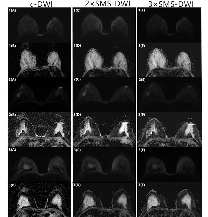
Fig1A~F(normal
breast),Fig2A~F(breast
carcinoma),Fig3A~F(fibroadenoma
of breast) Example of diffusion
weighted images and corresponding ADC maps acquired with conventional
EPI(c-DWI) as well as with two-fold(2×SMS-DWI) and
three-fold(3×SMS-DWI) simultaneous multi-slice acquisition.2×SMS-DWI showed
equivalent image quality as compared to c-DWI.Image quality significantly
deteriorated in 3×SMS-DWI.The ADC maps obtained in 2×SMS-DWI and 3×SMS-DWI
showed equivalent image quality as compared to c-DWI.