4317
TGSE diffusion-weighted pulse sequence in the evaluation of optic neuritis: A comprehensive comparison of image quality with RESOLVE DWI1Shanghai Insititute of Medical Imaging, Shanghai, China, 2Eye & ENT Hospital of Shanghai Medical School, Fudan University, Shanghai, China, 3Siemens Healthcare Ltd, Shanghai, China, 4Department of Radiology, Shanghai Ninth People’s Hospital, Shanghai JiaoTong University, Shanghai, China, 5Department of Radiology, Eye & ENT Hospital of Shanghai Medical School, Fudan University, Shanghai, China, 6Department of Digitalization, Siemens Shenzhen Magnetic Resonance Ltd., Shenzhen, China
Synopsis
This study investigated the role of RESOLVE and TGSE DWI sequences in the evaluation of optic neuritis and compared their image qualities qualitatively and quantitatively. We found that TGSE significantly improved the image quality for the evaluation of optic neuritis by reducing the susceptibility induced image distortion compared with RESOLVE. However, it appeared lower SNR and CNR than that of RESOLVE images.
Purpose:
To investigate and compare the role of readout segmented echo-planer imaging (RESOLVE) and a 2D turbo gradient and spin-echo (TGSE) diffusion-weighted (DW) pulse sequence with non-Cartesian BLADE trajectory in the evaluation of optic neuritis qualitatively and quantitatively.Materials and methods:
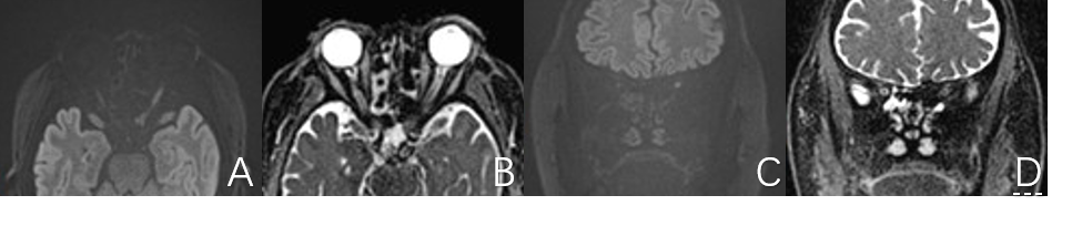
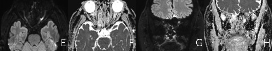
Both RESOLVE and TGSE images of 34 optic nerves were acquired from 28 patients on a 3T MR scanner (MAGNETOM Prisma, SIEMENS Healthcare, Erlangen, Germany) with a 64-channel head&neck coil. The parameters for TGSE BLADE DWI were as follows: TR/ TE = 4000/62 ms, slice thickness/gap = 2/0.2 mm, slices = 21, bandwidth = 520 Hz/Px, field of view (FOV) = 280 × 280 mm, matrix = 192 × 192, voxel size = 1.5 × 1.5 × 2.0 mm, number of excitations (NEX) = 1, 4 scan trace mode, b = 0, 1000 s/mm2, turbo factor = 13, EPI factor = 3, and the data acquisition time was 3min46s. For REOSLVE DWI, the imaging parameters were: TR/TE = 5020/53 ms, slice thickness/gap = 2/0.2 mm, slices = 21, bandwidth =766Hz/Px, field of view (FOV) = 230 × 230 mm, matrix = 192 × 192, voxel size = 1.2 × 1.2 × 2.0 mm, 4 scan trace mode, b = 0, 1000 s/mm2, and the data acquisition time was 3min46s. Both sequences were applied for transversal and coronal planes, respectively. Conspicuity and the distortions of lesions, degree of ghosting artifact, uniformity of fat suppression, and the overall image quality of the RESOLVE and TGSE images were qualitatively evaluated by two radiologists (scoring details seen in table 1). Distortion was also quantitatively evaluated by comparing the distances between the same anatomic points on TSE-T1WI, TSE-T2WI, RESOLVE and TGSE images. The apparent diffusion coefficient (ADC) values, signal-to-noise ratios (SNRs), and contrast-to-noise ratios (CNRs) of the two DWIs were compared as well.Results:
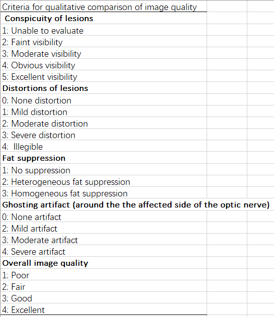
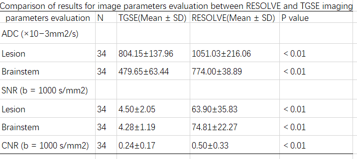
The comparisons of the qualitative scores indicated that TGSE significantly improved the uniformity of fat suppression and reduced degree of ghosting artifact. The distortion of the lesions is extremely improved, especially in ADC maps. However, the conspicuity of lesions is better on RESOLVE images. Quantitative evaluations revealed that TGSE almost completely remove the distortion. The ADC values of the optic nerves on TGSE were lower than those on RESOLVE, as well as the brainstem. Both of the SNR and CNR of TGSE were lower than RESOLVE. The results of the comparisons mentioned above are listed in table 2.Conclusion:
TGSE significantly improved the image quality for evaluations of optic neuritis by reducing the susceptibility artifacts and distortion compared with RESOLVE. However, it appeared blurrier than RESOLVE images.Acknowledgements
References
1. Hu Houchun H,McAllister Aaron S,Jin Ning et al. Comparison of 2D BLADE Turbo Gradient- and Spin-Echo and 2D Spin-Echo Echo-Planar Diffusion-Weighted Brain MRI at 3 T: Preliminary Experience in Children.[J] .Acad Radiol, 2019, undefined: undefined.
2. Held P,Seitz J,Fründ R et al. Comparison of two-dimensional gradient echo, turbo spin echo and two-dimensional turbo gradient spin echo sequences in MRI of the cervical spinal cord anatomy.[J] .Eur J Radiol, 2001, 38: 64-71.
3. Porter DA, Heidemann RM. High resolution diffusion-weighted imaging using readout-segmented echo-planar imaging, parallel imaging and a two-dimesional navigator-based reacquisition. Magn Reson Med. 2009;62:468-475.
4. De Foer B, Vercruysse JP, Pilet B, et al. Single-shot, turbo spin-echo, diffusion-weighted imaging versus spin-echo-planar, diffusion-weighted imaging in the detection of acquired middle ear cholesteatoma. AJNR. American journal of neuroradiology. Aug 2006;27(7):1480-1482.
5. Dhepnorrarat RC, Wood B, Rajan GP. Postoperative non-echo-planar diffusion-weighted magnetic resonance imaging changes after cholesteatoma surgery: implications for cholesteatoma screening. Otology & neurotology : official publication of the American Otological Society, American Neurotology Society [and] European Academy of Otology and Neurotology. Jan 2009;30(1):54-58.
Figures