4306
Improving X-PROP with a more stable echo train for diffusion weighted MRI1Neuroradiology, Barrow Neurological Institute, Phoenix, AZ, United States, 2Philips Healthcare, Gainesville, FL, United States
Synopsis
EPI-based DWI is widely used in the clinic but suffers from geometric distortions. DW-PROPELLER, based on FSE, is free from geometric distortions but has low scan efficiency. X-PROP was developed to improve FSE-based DW-PROPELLER scan efficiency by employing a GRASE readout. Although more efficient, XY2 phase modulation used in X-PROP is sensitive to the flip angles of the RF pulse train. This project improves X-PROP image quality by incorporating LRX phase modulation to increase SNR and signal stability. Image quality improvement was illustrated by comparing in vivo images produced with LRX phase modulation, XY2 phase modulation, and SPLICE PROPELLER imaging.
Introduction
Diffusion weighted (DW) MRI plays an important role in routine neuro imaging. DW EPI is widely used primarily because of its SNR efficiency but suffers from geometric distortions, especially in high resolution scans. Efforts have been made to reduce DW EPI image distortions by using very complicated calibration/correction approaches. 1-3 By contrast, FSE-based DW MRI, including PROPELLER, 4 has been shown to be relatively free of geometric distortions, but suffers from low scan efficiency. Turboprop, 5 X-PROP 6, and steer-PROP 7 were subsequently developed to improve scan efficiency. XY2 phase modulation 4 is used in both turboPROP and X-PROP to mitigate the violation of the CPMG condition, requiring high RF pulse train flip angles, resulting in fast signal decay and signal instability. LRX phase modulation was designed to produce higher and more stable signal quality, with reduced sensitivity to the flip angle variation. 8,9 In this work we incorporate LRX phase modulation into X-PROP to improve image quality, and then compare it with original XY2 phase modulation, as well as another emerging technique, SPLICE PROPELLER 10,11.Methods
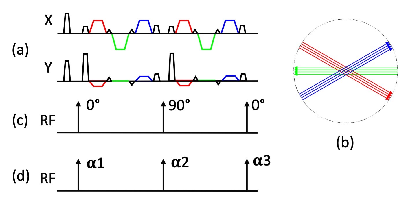
X-PROP adopts a GRASE readout to improve the data acquisition efficiency and places different gradient or spin echoes in distinct blades in k-space to mitigate artifacts caused by the susceptibility induced phase errors (Fig. 1a-b). 6 The previous XY2 phase modulation implementation of X-PROP alternates the RF refocusing pulses between X and Y axis (Fig. 1c), with odd and even echo data processed separately to avoid mixing data within the same blade. 4 LRX phase modulation applies a set of deliberately designed phases to the RF pulses (Fig. 1d), to improve signal stability and overall SNR. 8,9 This project incorporates LRX phase modulation into X-PROP on a 3T Philips Ingenia scanner (Philips Healthcare, Best, the Netherlands) with a 13 channel head coil. Data were acquired on volunteers with common imaging parameters: FOV = 240x240 mm2, res = 1.25x1.25 mm2, 24 4-mm slices, SENSE reduction factor R = 2, diffusion encoding on 3 orthogonal directions with b = 1000 s/mm2, linear or center-out (low-high) in-blade view order, PROPELLER Nyquist oversampling factor and #averages of b high data were adjusted for each scan to match scan time (~ 4 min). Additional parameters for X-PROP with both XY2 and LRX phase modulation include 1) ETI factor = 3, ETL = 16, b high average = 1; or 2) EPI factor = 5, ETL = 12, b high average = 2. Reference images were also acquired with SPLICE PROPELLER with ETL = 13, b high average = 1. Image reconstruction was done online on the host computer.Results and Discussions
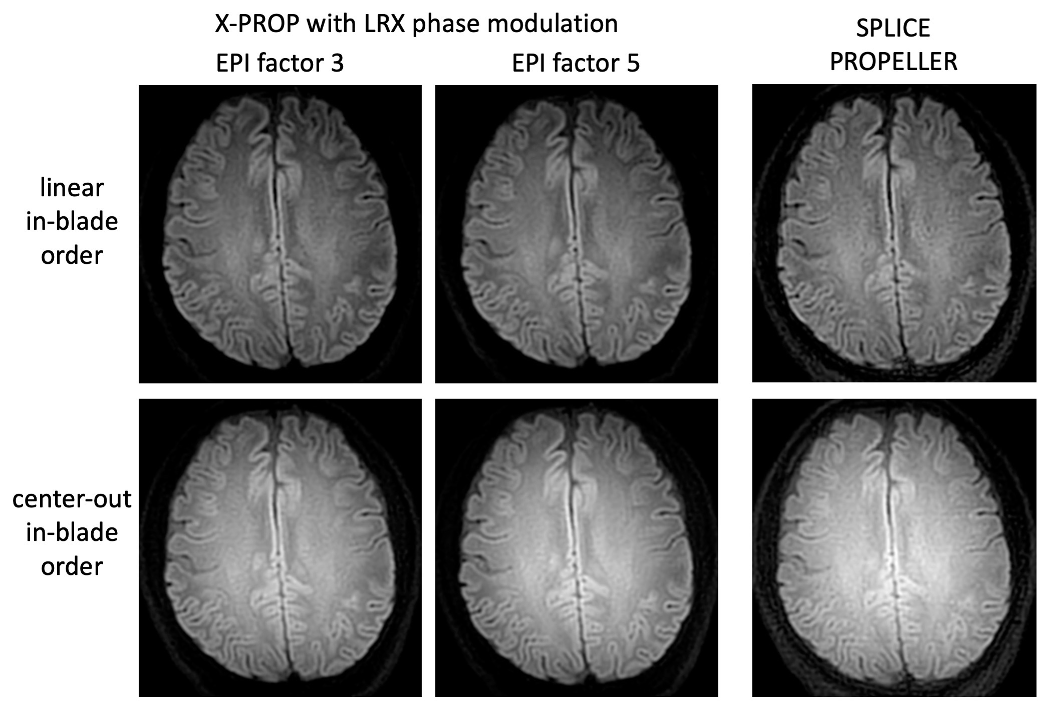
Fig. 2 shows improvement with LRX phase modulation over original XY2 phase modulation. Both data sets were acquired with EPI factor = 5 and linear in-blade view order. LRX phase modulation images demonstrate improved SNR but slightly reduced contrast compared with images obtained with XY2 phase modulation. Subtly reduced T2 contrast is likely due to reduction of the fast signal decay associated with XY2 phase modulation. This reduced T2 contrast with LRX phase modulation may help alleviate the T2 shine-through effect.In Fig. 3, X-PROP images with LRX phase modulation acquired with EPI factor of 3 and 5 are compared to SPLICE PROPELLER, all with linear in-blade view order. Both X-PROP data sets exhibit higher SNR than SPLICE PROPELLER. Although susceptibility induced artifacts are present in X-PROP data, they remain minimal when compared to those typically seen with EPI (not shown here). In this experiment, the PROPELLER Nyquist oversampling factor and # averages of b high data are 1.625/1 and 1.875/2 for SPLICE PROPELLER and X-PROP with an EPI factor of 5, respectively. For this imaging protocol (including FOV and resolution), the shortest scan time of X-PROP with an EPI factor of 5 is 1:58, about 2 times faster than SPLICE PROPELLER.
Fig. 4 shows the improvement in SNR using a center-out in-blade view order compared to the typical linear view order, with slightly reduced T2 contrast (and therefore less T2 shine-through effect) due to the shortened TE. The combination of increased SNR and reduced T2 shine-through effect may help improve diffusion weighting.
Conclusion
X-PROP imaging using LRX RF phase modulation demonstrates improved image quality compared to X-PROP using XY2 phase modulation and SPLICE PROPELLER. Therefore, X-PROP with LRX phase modulation provides a promising tool for fast and distortion-free DW MRI.Acknowledgements
This work is partially supported by Philips Healthcare.References
1. Liao C, et al. High-fidelity, high-isotropic-resolution diffusion imaging through gSlider acquisition with B1+ and T1 corrections and integrated DB0/Rx shim array. Magn Reson Med 2020;83:56-67.
2. Dong Z, et al. Tilted-CAIPI for highly accelerated distortion-free EPI with point spread function (PSF) encoding. Magn Reson Med 2019;81:377-392.
3. In MH, et al. Distortion-free imaging: A double encoding method (DIADEM) combined with multiband imaging for rapid distortion-free high-resolution diffusion imaging on a compact 3T with high-performance gradients. J Magn Reson Imaging 2019. DOI.org/10.1002/jmri.26792.
4. Pipe JG, et al. Multishot diffusion weighted FSE using PROPELLER MRI. Magn Reson Med 2002;47:42-52.
5. Pipe JG, et al. Turboprop: Improved PROPELLER imaging. Magn Reson Med 2006;55:380-385.
6. Li Z, et al X-PROP: A fast and robust diffusion-weighted PROPELLER technique. Magn Reson Med 2011;66:341-347.
7. Srinivasan G, Rangwala N, Zhou XJ. Steer-PROP: a GRASE-PROPELLER Sequence with Interecho Steering Gradient Pulses. Magn Reson Med. 2018;79;2533-2541.
8. Le Roux P. Non-CPMG fast spin echo with full signal. J Magn Reson 2002;155:278-292.
9. Li Z, et al. Robust diffusion weighted imaging using split-blade PROPELLER and LRX phase modulation. ISMRM 2009;p3516.
10. Deng J, Omary RA, Larson AC. Multishot Diffusion-Weighted SPLICE PROPELLER MRI of the Abdomen. Magn Reson Med. 2008;59:947-953.
11. Zhao X, Li Z, Gaddipati A. PROPELLER DUO: Applied to Diffusion-Weighted Imaging. In proceedings of ISMRM, 2009, p3518, Hawaii, USA.
Figures