4302
Robust Diffusion-Weighted Imaging near Metallic Objects with Inner-FOV Single-Shot STEAM based on 2D-Selective RF Excitations1Department of Systems Neuroscience, University Medical Center Hamburg-Eppendorf, Hamburg, Germany
Synopsis
The potential of a single-shot stimulated echo acquisition mode (STEAM) sequence based on RF refocused echoes for DW imaging close to metallic objects is evaluated. It is optimized for spinal cord applications by combining it with inner-FOV technique based on 2D-selective RF (2DRF) excitations and half-Fourier sampling which improves its signal-to-noise ratio (SNR) efficiency significantly. Its robustness in the presence of metallic objects is investigated and compared to EPI showing a better performance with smaller regions suffering from signal losses.
Introduction
Diffusion weighted (DW) imaging is a powerful tool to access white matter fibre tracts and tissue microstructure and could be valuable to characterize and/or monitor spinal cord injuries. While inner field-of-view (FOV) echo-planar imaging (EPI) is able to provide a good image quality for DW imaging of the spinal cord1,2, it suffers from severe artifacts in the presence of metallic objects making it unusable in regions close to implants stabilizing the lower spine. Here, the potential of a single-shot stimulated echo acquisition mode (STEAM) sequence3 based on RF refocused echoes for DW imaging close to metallic objects is evaluated. It is optimized for spinal cord applications by combining it with inner-FOV technique based on 2D-selective RF (2DRF) excitations4 and half-Fourier sampling5 which improves its signal-to-noise ratio (SNR) efficiency significantly. Its robustness in the presence of metallic objects is investigated and compared to EPI showing a better performance with smaller regions suffering from signal losses.Methods
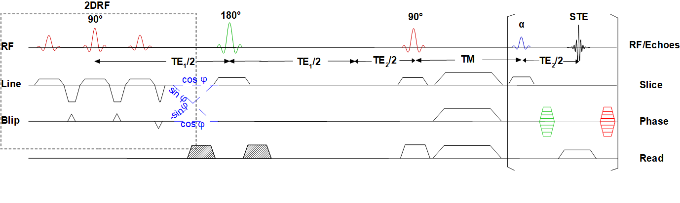
The basic pulse sequence used in this study is presented in Figure 1. It is based on an initial 2DRF excitation with a blipped-planar trajectory tilted by an angle ϕ compared to the slice and phase-encoding direction in order to shift the unwanted side excitations out of the refocussing plane and the slice stack to be measured which reduces the field-of-excitation (FOE) and the RF pulse length considerably2. The 2DRF envelopes were calculated using the low-flip-angle approximation4 and designed to excite a rectangular profile with a size of 4×56mm² (slice×phase-encoding direction) using a trajectory with a resolution of 4.0×10.0mm² in the line×blip direction.Single-shot STEAM and EPI Images were acquired on a 3T whole-body MR system (PrismaFit, Siemens) using a 64-channel head-neck coil using a voxel size of 1.0x1.0x4.0 mm³ and covering either a full FOV of 100x128 mm² or an inner FOV of 32x128 mm². The readout flip angle of single-shot STEAM was adapted to yield a point-spread function with a full-width at half-maximum (FWHM) corresponding to the nominal resolution3. For diffusion tensor imaging (DTI), 20 slices with 8 averages of 6 diffusion directions with a diffusion weighting of 500 s mm-2 and a non-diffusion weighted image were acquired. TR was between 4000 and 8000 ms resulting in total acquisition times between 6.3 and 11.2 min.
A water phantom and a cucumber larded with 20-cent coins (€) as metallic objects and healthy volunteers were investigated, the latter giving their informed consent prior to examination.
Results and Discussion
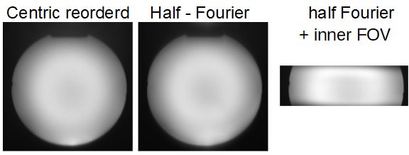
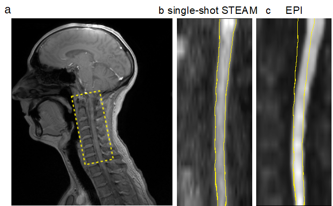
Results and Discussion (220) Figure 2 shows the advantages of half-Fourier sampling and inner-FOV acquisition for single-shot STEAM. With both techniques applied, the acquisition time is reduced from 1183 ms to 264 ms while there is almost no SNR penalty yielding an overall increase of the SNR efficiency by about 100%, While both techniques reduce the number of k-space lines and, thus, decrease the SNR at a first glance, the modified sampling allows for larger readout flip angles (e.g. from 12° to 22°) for the same FWHM of the point-spread function which increases the signal amplitude.Results of in vivo acquisitions are presented in Figure 3. While EPI is much faster (6.2 min vs 11.2 for single-shot STEAM) and provides a better SNR, it suffers from geometric distortions caused by global and local field inhomogeneities even in the absence of metallic objects. In contrast, single-shot STEAM is more robust and, e.g., does not show a modulation of the posterior edge along the spinal cord.
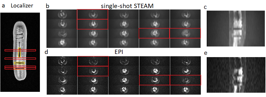
This is demonstrated in Fig. 4 showing the results of experiments in the phantom with metallic objects. EPI not only suffers from pronounced signal loses in slices containing parts of the coins, but also in neighbouring sections. On the other hand, single-shot STEAM is rather robust, even in regions close to the coins, and clearly shows the exact position and shape of the coins.
Conclusion
The SNR efficiency of single-shot STEAM can be significantly improved by using inner-FOV techniques, e.g. based on 2DRF excitations, and half-Fourier sampling. Compared to EPI it still suffers from a reduced SNR but is much less prone to metallic objects and field inhomogeneities. Thus, it could be a viable technique to investigate spinal cord regions close to implants.Acknowledgements
This work was supported by a grant from Wings for Life.References
1. Saritas EU, Cunningham CH, Lee HJ, et al., DWI of the spinal cord with reduced FOV single-shot EPI. MRM, 2008;60: 468-473
2. Finsterbusch J,Improving the performance of diffusion-weighted inner field-of-view echo-planar imaging based on 2D-selective radiofrequency excitations by tilting the excitation plane. JMRI, 2012;35: 984-992
3. Nolte UG, Finsterbusch J, Frahm J, Rapid Isotropic and Diffusion Mapping and Without Susceptibility and Artifacts: Whole and Brain Studies and Using Diffusion-Weighted and Single-Shot STEAM and MR Imaging. MRM, 2000;
4. Pauly J, Nishimura D, Macovski A, A k-Space Analysis and of Small-Tip-Angle and Excitation. JMR. 1989;81:4
5. Margosian P, Schmitt F, Purdy DE, Faster MR imaging: Imaging with half the date. Health Care Instrum. 1994;32: 535-539
Figures