4297
SNR efficient diffusion imaging at 7T with B1+ mitigated multi-shot SMS-EPI, using semi adiabatic PINS RF and low-rank completion reconstruction1Center for Neuroscience Imaging Research, Institute for Basic Science (IBS), Suwon, Korea, Republic of, 2Department of Computer Science, Mathematics, Physics, and Statistics, University of British Columbia, Kelowna, BC, Canada, 3Athinoula A. Martinos Center for Biomedical Imaging, Massachusetts General Hospital, Charlestown, MA, United States, 4Department of Radiology, Harvard Medical School, Boston, MA, United States, 5Biomedical Engineering and Imaging Institute, Icahn School of Medicine at Mount Sinai, New York, NY, United States
Synopsis
B1+ inhomogeneity, SAR, and shortened T2-relaxation are the main challenges to leverage the higher SNR at ultra-high-field MRI. Here, we develop a new method by combining navigation-free multi-shot SMS-EPI with low-rank matrix completion reconstruction with semi-adiabatic PINS pulse for B1+ insensitive SMS imaging. Using this combined approach, we demonstrated mitigated B1+ inhomogeneity by comparing with conventional-SE pulse and the feasibility of low-rank completion reconstruction at high b-value. Finally, 1.2 mm isotropic whole-brain diffusion MRI was acquired across 64 diffusion directions with high-SNR in 11 minutes at 7T.
Introduction
Ultra-high-field (UHF) MRI offers the opportunity for higher spatial resolution imaging from increased SNR1-2. However, the increased B1+ inhomogeneity3, SAR4, and shortened T2-relaxation pose challenges for practical applications, particularly for diffusion MRI (dMRI). Advanced diffusion imaging strategies, including 3D-multislab acquisition with non-linear slab-profile encoding5 and the use of parallel transmission6, have been proposed to mitigate some of these issues to allow high-quality, high-resolution dMRI. In this work, we develop a new SNR-efficient dMRI approach for UHF to tackle the aforementioned issues. Our approach incorporates: i) navigation-free multi-shot SMS-EPI with low-rank matrix completion reconstruction (MUSSELS)7-9 and ii) A SEmi-Adiabatic Matched-phase Spin echo (SEAMS) PINS Pulse-pair for B1+ insensitive SMS imaging10. The use of multi-shot SMS-EPI will allow high MB and Rinplane accelerations (e.g. Rinplane×MB = 3×7), which help achieve short-TR for high SNR-efficiency and short-TE to reduced signal loss from shortened T2. The high MB factor also work synergistically with the SEAMS-PINS pulse design, which is more favorable at high MB factors, to enable B1+ mitigated low-SAR spin-echo acquisition. Using this combined approach, 1.2 mm isotropic whole-brain dMRI was acquired across 64 diffusion directions with high-SNR in 11 minutes.Methods
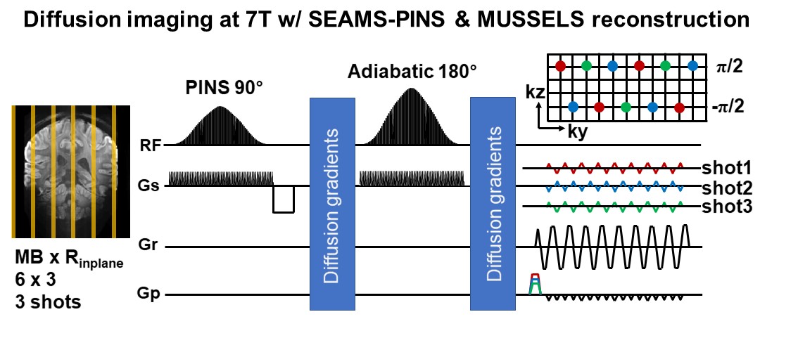
The sequence diagram for the proposed multi-shot SMS-EPI dMRI is shown in Figure 1. In this exemplar sagittal acquisition case at 1.2 mm isotropic resolution, data were acquired across 3 EPI-shots, each at an acceleration factor of Rinplane×MB = 3×6 (with an effective 6-fold acceleration across 3 shots). To enable good MUSSELS reconstruction, k-space sampling was performed to provide FOV/6 CAIPI-shift for each EPI-shot and FOV/2 CAIPI-shift for the merged data (right side of Figure 1). The SEAMS-PINS pulses were designed to obtain a 1/19 slice separation ratio for 1.2 mm slice thickness, resulting in a slice separation of 22.8mm. Adiabatic Shinnar Le-Roux (SLR) algorithm11 was employed to create the 180° pulse with a duration of 11.2ms and the matched-phase 90° pulse with a duration of 21.3ms.Data were collected from three healthy subjects after obtaining informed consent using a 7T MRI scanner (Siemens MAGNETOM Terra 7T, Siemens, Erlangen), equipped with a single-channel transmitter, and a 32-channel receive head coil (Nova Medical, Wilmington, MA). Scans were performed on two subjects to compare B1+ inhomogeneity effects between SEAMS-PINS and conventional-SE pulses. The feasibility of the MUSSELS reconstruction in the proposed sequence was also examined by comparing against a standard SENSE reconstruction. The imaging parameters were: 1.2 mm isotropic sagittal acquisition, Rinplane×MB = 3×6, 3-shots, FOVxy = 180×180mm2, 114 slices, p.f.=6/8, resolution, TR/TE = 6000/58ms (TR limited by high-SAR in conventional-SE case). Additionally, in one subject, 1.2 mm isotropic diffusion data were acquired across 64 diffusion directions along with 4 b=0 acquisitions, resulting in a total scan time of ~11 minutes. The same imaging parameters as above were used, with an increase in MB factor to 7 to enlarge the FOV coverage along the L-R direction for this subject. Data were acquired at b = 1000s/mm2 with a TR/TE of 3000/62ms. Note: a TR as short as 2s is achievable with this sequence while staying within the SAR-limits. However, a TR of 3s was chosen as it already provides an SNR-efficiency of >90%, while being much less sensitive to spin-history effects from motion12.Results
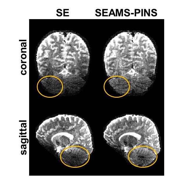
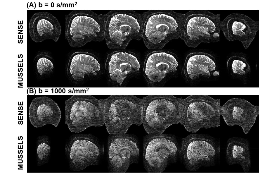
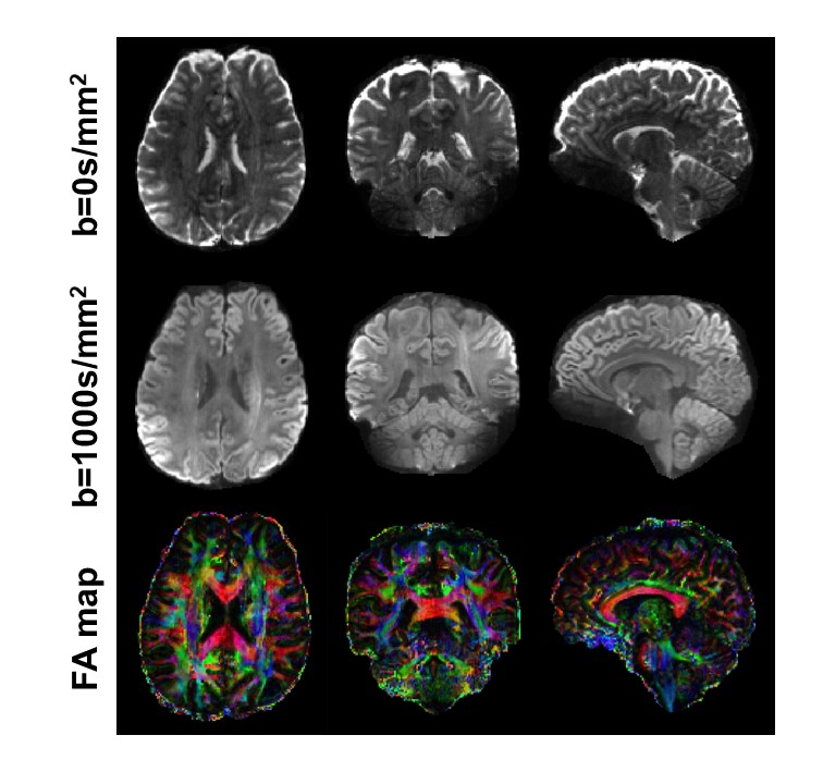
Figure 2 shows MUSSELS reconstructed images from conventional-SE and SEAMS-PINS pulses respectively. The images from conventional-SE show severe signal dropout due to B1+ inhomogeneity in the cerebellum region, while those acquired with SEAMS-PINS are seen to be more robust. The results of the MUSSELS reconstruction on the 3-shot DW acquisition were compared against those from a conventional SENSE reconstruction in Figure 3 for a particular SMS slice group. The artifacts arising from diffusion phase corruption between EPI-shots are apparent in the SENSE reconstruction, while the MUSSELS reconstruction successfully mitigate these artifacts. Figure 4 shows b=0 and averaged DWI images, along with the corresponding FA map in axial/coronal/sagittal views, where high-SNR results were obtained at 1.2 mm isotropic whole-brain.Discussion and Conclusion
We have proposed a multi-shot SMS-EPI dMRI approach for UHF that utilizes SEAMS-PINS pulse and MUSSELS reconstruction. This combined approach was developed to mitigate the challenges from increased B1+ inhomogeneity, SAR, and shortened T2-relaxation at UHF. We demonstrated that such acquisition to allow high SNR-efficiency dMRI across the whole-brain at high resolution. We aim to further incorporate new advancement in multi-shot SMS-EPI reconstruction using the SMS-NEATER9 approach, which should further refine our reconstruction and push forward acceleration capability.Acknowledgements
This work was supported by IBS-R015-D1, NIH R01EB019437, R01 MH116173, and U01EB025162.References
[1] Ugurbil K, Adriany G, Andersen P, et al. Ultrahigh field magnetic resonance imaging and spectroscopy. Magn Reson Imaging 2003; 21:1263–1281.[2] Duyn JH. The future of ultra-high field MRI and fMRI for study of the human brain. Neuroimage 2012; 62:1241–1248.[3] Van de Moortele PF, Akgun C, Adriany G et al. B1 destructive interferences and spatial phase patterns at 7 T with a head transceiver array coil. Magn. Reson. Med. 2005; 54(6): 1503–1518.[4] Hoult DI, Phil D. Sensitivity and power deposition in a high-field imaging experiment. J. Magn. Reson. Imaging 2000; 12(1): 46–67.[5] Wu W, Poser BA, Douaud G et al. High-resolution diffusion MRI at 7T using a three-dimensional multi-slab acquisition. NeuroImage 2016; 143: 1–14.[6] Wu X, Auerbach EJ, Vu AT et al. High-resolution whole-brain diffusion MRI at 7T using radiofrequency parallel transmission. Magn Reason Med 2018; 80:1857–1870.[7] Mani M, Jacob M, Kelley D et al. Multi-shot sensitivity-encoded diffusion data recovery using structured low-rank matrix completion (MUSSELS). Magn. Reson. Imaging 2017; 78:494–507.[8] Mani M, Jacob M, McKinnon G et al. SMS MUSSELS: A navigator-free reconstruction for simultaneous multi-slice-accelerated multi-shot diffusion weighted imaging. Magn Reason Med 2020; 83:154–169.[9] Bilgic B, Chatnuntawech I, Manhard MK et al. Highly accelerated multishot echo planar imaging through synergistic machine learning and joint reconstruction. Magn Reason Med 2019; 82:1343–1358.[10] Feldman RE, Islam HM, Xu J et al. A SEmi-Adiabatic Matched-Phase Spin Echo (SEAMS) PINS pulse-pair for B1-insensitive simultaneous multislice imaging. Magn. Reson. Med. 2016; 75:709–717.[11] Pauly J, Le Roux P, Nishimura D, Macovski A. Parameter relations for the Shinnar-Le Roux selective excitation pulse design algorithm [NMR imaging]. IEEE Trans Med Imaging 1991; 10:53–65.[12] Engstrom M, Martensson M, Avventi E et al. On the signal-to-noise ratio efficiency and slab-banding artifacts in three-dimensional multislab diffusion-weighted echo-planar imaging. Magn. Reson. Med. 2015; 73:718–725.
Figures