4281
3D Surface Scanning an MRI Machine: Initial Experience and Preliminary Results
Nicole Wake1,2, Maggie Fung3, Stephen Dashnaw4, J. Thomas Vaughan Jr.4, and Fraser Robb5
1Department of Radiology, Montefiore Medical Center, Albert Einstein College of Medicine, Bronx, NY, United States, 2Center for Advanced Imaging Innovation and Research, Department of Radiology, NYU School of Medicine, New York, NY, United States, 3GE Healthcare, New York, NY, United States, 4Columbia University, New York, NY, United States, 5GE Healthcare, Aurora, OH, United States
1Department of Radiology, Montefiore Medical Center, Albert Einstein College of Medicine, Bronx, NY, United States, 2Center for Advanced Imaging Innovation and Research, Department of Radiology, NYU School of Medicine, New York, NY, United States, 3GE Healthcare, New York, NY, United States, 4Columbia University, New York, NY, United States, 5GE Healthcare, Aurora, OH, United States
Synopsis
An approach to 3D capture a life-sized MRI machine using 3D surface scanning is described. The 3D files generated can be used for 3D printing, augmented or virtual reality, or simulation studies.
Introduction
Three-dimensional (3D) surface scanning is a non-invasive method to capture the shape, texture, and volume information of a 3D object. In medicine, 3D surface scanning is generally used to capture the 3D geometry information of real patient anatomy,1-3 but it can also be used to get 3D information regarding medical devices and equipment. Surface scanning technologies rely on different physical principles which all work to reconstruct an object in slightly different ways. Categories include laser triangulation 3D scanning which projects a laser beam on a surface and measures the deformation of the laser ray; structured light 3D scanning which measures the 3D shape of an object using projected light patterns and a camera system; photogrammetry which involves the use of cameras that take photos in a range of positions, angles and distances from the object; and contact-based 3D scanning which relies on the sampling of several points on a surface measured by the deformation of a probe. The result is a 3D file of the object which can be saved in a computer aided design (CAD) file format that can be edited, visualized in augmented reality (AR) or virtual reality (VR), 3D printed, or used in simulations. The objective of this work was to determine if it was possible to capture the 3D geometry of a magnetic resonance imaging (MRI) system using 3D scanning techniques.Methods
To 3D capture an MRI machine, a 3 Tesla MRI system (Premier, GE Healthcare, Waukesha, WI) was surface scanned before it was ramped up. Surface scanning was performed with a Leica RTC360 Laser Scanner (Leica Geosystems, St. Gallen, Switzerland) and an Artec Eva hand-held scanner (Artec3D, Luxembourg) [Figure 1]. The Leica RTC360 laser scanner was placed in 8 positions around the scan room [Figure 2] and the hand-held scanner was used to add in information regarding the table and scanner bore.Results
3D surface scanning was successfully carried out with both 3D scanning methods. To generate the final 3D MRI CAD file, 3D image post-processing was performed using several different software packages including: Leica Cyclone Register 360 (Leica Geosystems, St. Gallen, Switzerland) for RTC scan alignment, Artec Studio 14 (Artec3D, Luxembourg) for Eva scan data processing, Rhino with plugin (Robert McNeel & Associates, Seattle, WA) for CAD modeling specific elements, Polyworks Modeler (Innovmetric, Quebec, QC, Canada), and Geomagic Wrap (3D Systems, Rock Hill, SC) for additional mesh cleanup. After significant post-processing, the final MRI model demonstrated a good visual match to the actual MRI system [Figure 3].Discussion and Conclusions
3D surface scanning is a useful method to capture the 3D geometry of medical equipment. The realistic and accurate 3D reconstructions obtained from 3D surface scanning can be used for many applications including 3D printing, AR/VR, or simulations. Specifically, the 3D reconstructed MRI model could be highly useful for developing improved designs for MR spaces and for allowing patients to tour an MRI suite prior to their imaging exam. Future studies will utilize the CAD model generated here to improve trainee education and to enhance the patient experience.Acknowledgements
The authors thank BluEdge (New York, NY) and Direct Dimensions, Inc (Owings Mills, MD) for assistance with 3D scanning and post-processing.References
1. Jones PRM et al. Three-Dimensional Surface Anthropometry: Applications to the Human Body. Optics and Lasers in Eng. Sept 1997, pp89-117.
2. Da Silveira et al. Craniofacial Applications of Three-Dimensional Laser Surface Scanning. Journal of Craniofacial Surgery. July 203, 14(4), p449-456.
3. Camison L et al. Validation of the Vectra H1 Portable Three-Dimensional Photogrammetry System for Facial Imaging. Int J Oral Maxillofac Surg. 2018 Mar; 47(3): 403-410.
Figures

Figure 1: Leica RTC360 laser scanner in position to capture an
image of the MRI system (left) and Artec Eva hand-held system shown scanning MR
bore (right).
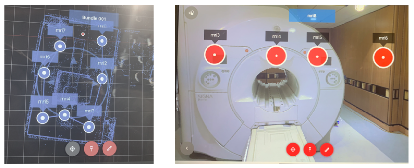
Figure 2: Map showing locations of Leica
TR360 scanner (left) and scanner positions overlaid onto photograph of MRI
system (right).
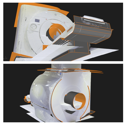
Figure 3: Front (top)
and back (bottom) views of the 3D reconstructed MRI system generated from 3D
scanning technologies.