4266
B1+ dependency on frequency in four human organs in three body models for 23Na, 13C and 31P whole-body birdcages at 7T.
Ria Forner1, Janot P. Tokaya2, Alexander Raaijmakers2,3, and Dennis Klomp2
1Radiology, UMC Utrecht, Utrecht, Netherlands, 2UMC Utrecht, Utrecht, Netherlands, 3Biomedical Engineering, Eindhoven University of Technology, Utrecht, Netherlands
1Radiology, UMC Utrecht, Utrecht, Netherlands, 2UMC Utrecht, Utrecht, Netherlands, 3Biomedical Engineering, Eindhoven University of Technology, Utrecht, Netherlands
Synopsis
This work is presenting a simulation study on X-nuclei imaging at 7T with a birdcage body coil for sodium, carbon and phosphorous. Three models and four imaging targets have been investigated. Although B1+ efficiency generally decreases with frequency, we show that the SAR efficiency, ( B1+/√SAR ) stays more or less constant over the investigated frequencies. Furthermore, it is shown that although every model and every imaging target has one optimal relative phase setting between the birdcage ports, for each frequency there exists one generic phase setting that provides only a minimal drop in performance over all imaging targets and models.
INTRODUCTION
At clinically relevant field strengths such as 1.5T or 3T as opposed to 7T and beyond, proton imaging makes use of the birdcage body coil as a general, large-field-of-view transmit coil and reception is performed using a local receive array. The wavelength of X-nuclear B1 fields is much larger and so, for these nuclei, the birdcage body coil can still be used to provide a relatively uniform excitation at ultra-high field.In this abstract, birdcage body coils tuned to 120MHz for 31P, 79MHz for 23Na, and 75 MHz for 13C at 7T were simulated with a human male, female or child body model are presented. The resulting B1+ and SAR efficiencies in four organs were extracted for comparison: the heart, the liver, the spleen and the kidneys. We have determined these metrics for the optimal phase settings of the birdcage feed ports and for a generic phase setting that aims to cover all investigated models and/or imaging targets.
METHODS
All simulations are performed using the numerical FDTD simulation package Sim4Life (ZMT, Zurich, Switzerland).The coil has an inner diameter of 60 cm, 24 rods at lengths of 40 cm, and an RF shield with 68 cm diameter. Lumped element capacitors with parallel resistive losses were used with realistic Q values to mimic coil losses. Three human body models (Duke, Ella, and Thelonius, an adolescent male) were each inserted coaxially inside the three high-pass birdcages (figure 1).
The power was normalised to 1W total input power to assume a perfectly matched coil with no reflections at the ports. In post-processing a final check of circular polarisation was performed by plotting the B-field vectors so as to visualise polarisation. This was performed to obtain optimal settings for all four organs, in all three body models and at all three frequencies. During post-processing, the phase of the second port was stepped from 0° through 360°to ascertain the optimum (for B1+) phase shim for each organ. Further, an average phase setting was determined for each frequency and the resulting loss in efficiency was determined when compared to the optimal phase setting for each specific case of the organs. Gridding was manually adjusted to provide a minimum voxel size of 2 mm3. The coil was driven in two ports separately using a CW power source matched to the coil port impedance and the fields summed using equal weighting. 3D B1+and SAR maps in the body for a length of 10cm beyond the end rings of the coil were exported to MATLAB (information about company and location of company) for analysis. Results were analysed by calculating the mean and standard deviation of B1+ in each target organ.
RESULTS
B1+ and SAR efficiency have been determined in each model, organ and frequency. Results are presented in figures 2 and 3. Figure 4 shows the sinusoidal behaviour of the mean B1+ for all organs, body models and the lowest and highest frequencies as the phase difference between the two ports is varied from 0° through 360°. Although the optimal phase settings vary, one generic phase setting can be identified for each frequency at which the penalty shouldn't be too large. Finally, in figure 5 we see that this penalty for deviating from the best shim setting is insignificant. At maximum, B1+ efficiency is reduced by 10%.DISCUSSION
The behaviour of the B1+ field follows similar trends at 75MHz and at 79MHz, although markedly different at 120MHz. As expected, at 120MHz, the mean B1+ is about half of that at the lower frequencies. This can be attributed to the fact that the tissue loading is increased at the higher frequency and thus, less field penetrates the body models. However, this trend disappears when the B1+ field is normalised by the square root of the peak local SAR. As the optimal phase shim is approached, the B1+ mean approaches the maximum while the homogeneity is also the highest. This is in accordance with the fact that a circularly polarised field is achieved in most of the organ of interest. The optimal shim settings for each organ in every body model may differ by as much as 70° (at 31P , for Duke, 30° for heart and 100° for spleen) This may be explained by the inhomogeneous field distribution in the body due to variable loading as well as the locations of the organs. It is possible to obtain a trade-off value for this phase shim setting at every frequency such that the performance is degraded by only between 5% to 10% irrespective of organ or subject.CONCLUSION
Although the B1+ decreases with frequency, the birdcage SAR efficiency stays more or less stable throughout the investigated frequency range (75MHz through 120MHz). It is possible to ‘shim’ the birdcage to obtain the maximum possible B1+ in every organ in a body. However, these shim settings vary widely according to organ and frequency and are very different from those used in clinical systems currently at similar frequencies. Still it is possible to determine a single optimal phase setting at every frequency such that the mean B1+ in every organ is hardly reduced compared to the maximum possible B1+.Acknowledgements
This work was supported by: European H2020-FETOPEN: NICI
References
No reference found.Figures
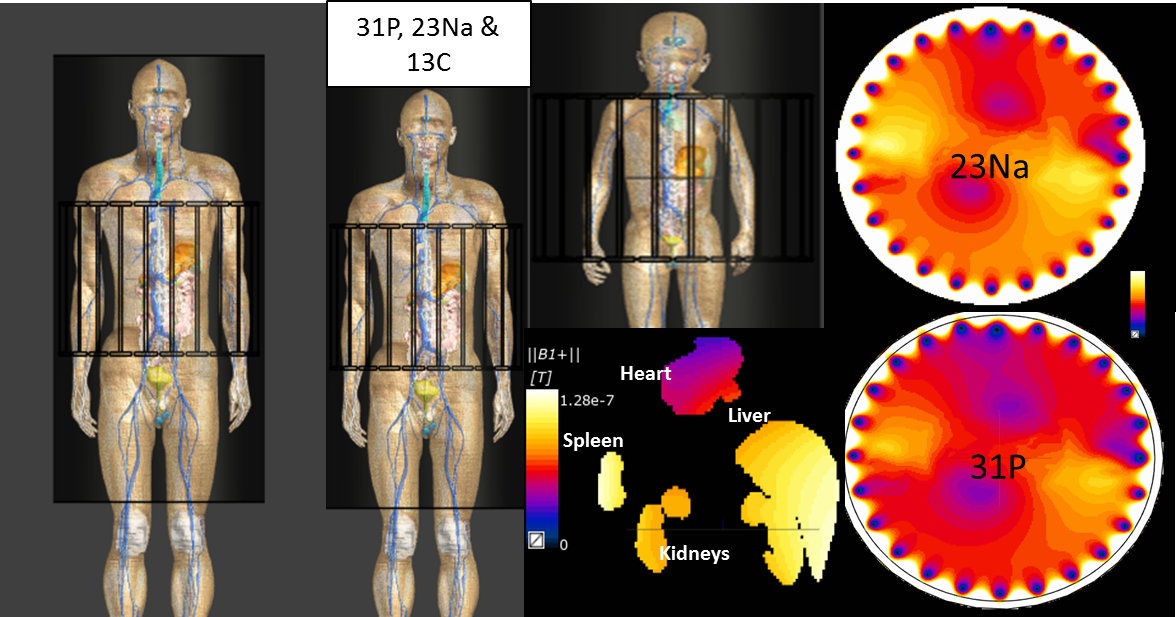
Figure 1: Clockwise from left to right; Configuration of setup with the three body models, Duke (adult male);
Ella (adult female) and Theolonius (adolescent male) in the birdcages; transverse slice through
iso-centre of Duke at 79MHz and 120MHz showing the variation in B1+ with
frequency; and B1+ in kidneys, heart liver and spleen in Duke at 31P
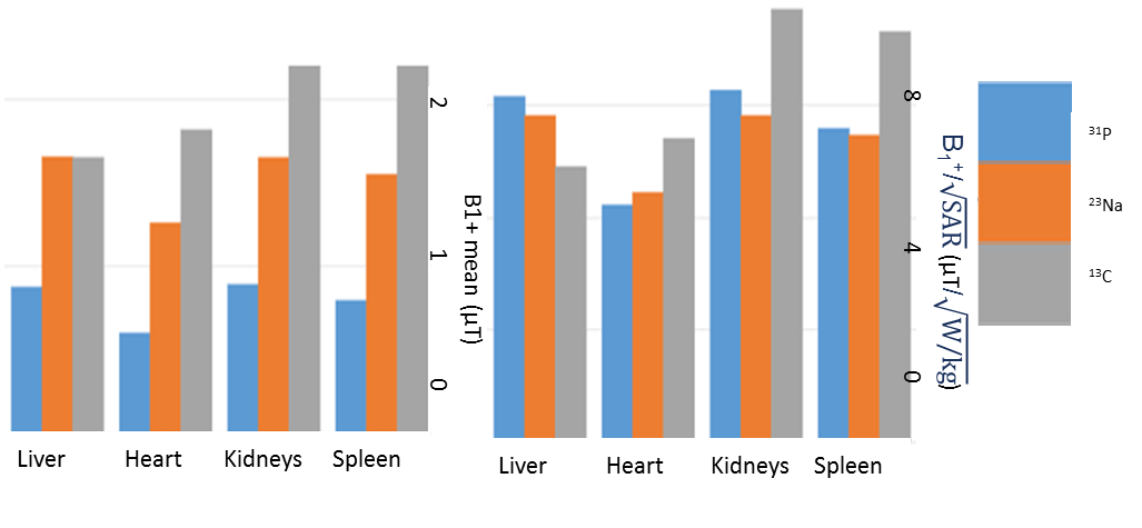
Figure 2: Simulations results comparing the trends in the mean B1+ efficiency and
the SAR efficiency in Duke at three frequencies and in four organs.
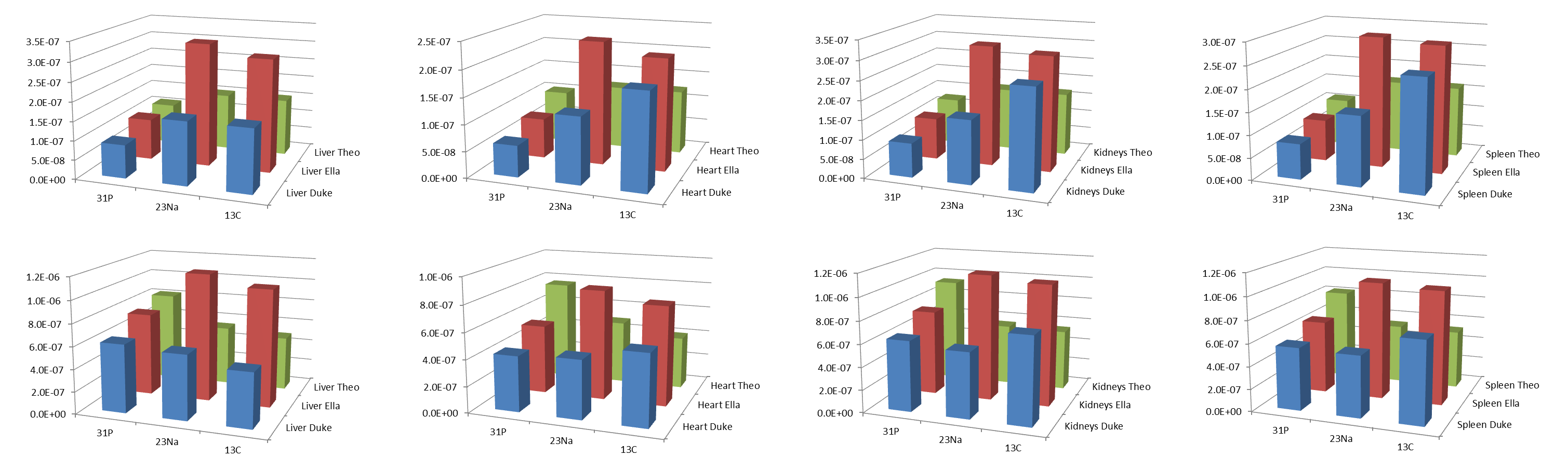
Figure 3: Simulations results comparing the trends in the mean B1+ efficiency (top row) and
the SAR efficiency (bottom row) in all three models at three frequencies and in four organs.
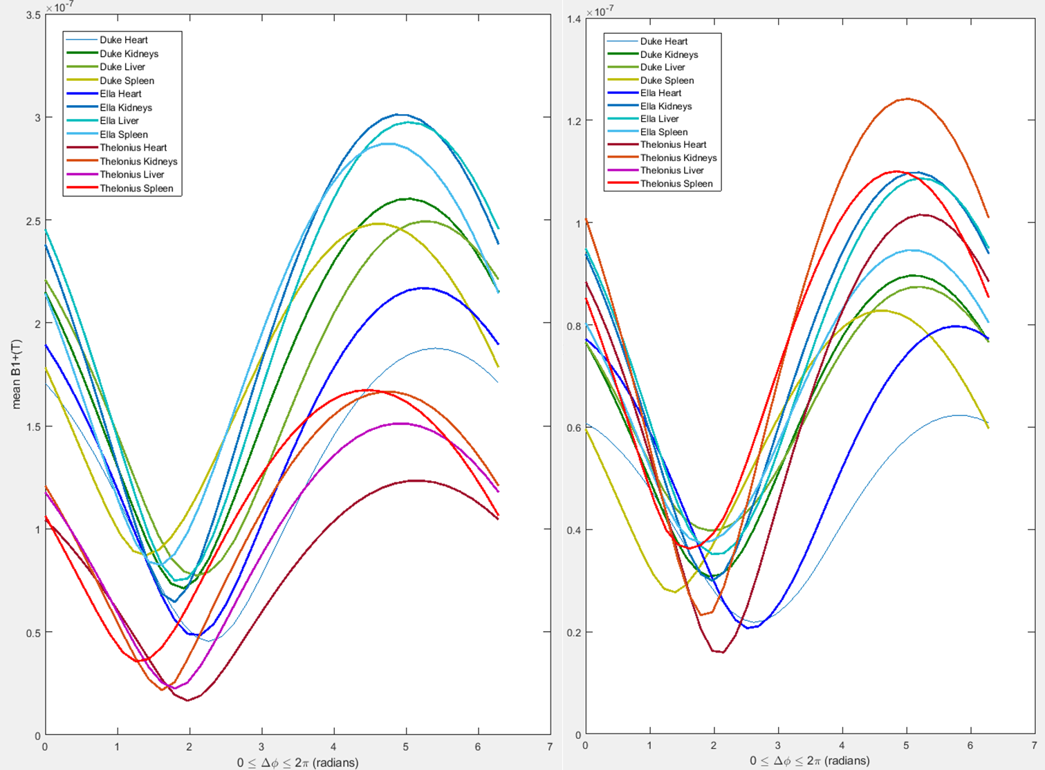
Figure 4: Effect of varying phase difference between
birdcage ports on the mean B1+ field as well as
homogeneity of B1+ field in the liver in Duke (green)
Ella (blue) and Thelonius (red)at 13C (left) and 31P (right).
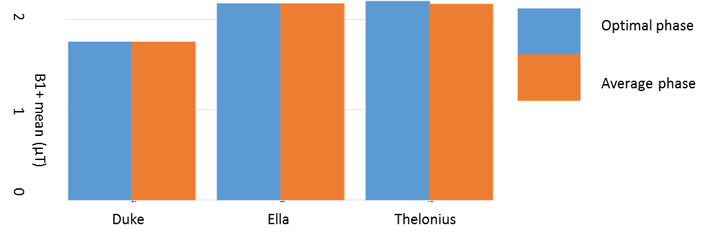
Figure 5: Insignificant difference between mean B1+ in liver
at 31P (shortest wavelength) in three body models with optimal shim
and average shim.