4233
Understanding the axial crushing effects of numerous current paths at 7 Tesla1Department of Radiology, University Medical Center Utrecht, Utrecht, Netherlands, 2Department of Biomedical Engineering, Eindhoven University of Technology, Eindhoven, Netherlands
Synopsis
MR spectroscopic images are often distorted by lipid signals in the skull. A crusher coil that generates a local magnetic field can suppress these signals, but a homogeneous crushing field is required. This work focusses on a solution for a homogeneous crushing field in the axial direction. It is shown through numerous tests with current patterns that wires circling the coil without any directioal component in the z-direction cause the most homogeneous crushing pattern. Currents in the z-direction should be compensated by a return current that cancels the magnetic field of the current.
Introduction
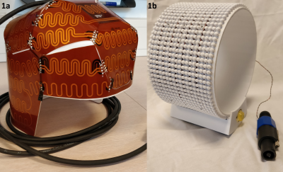
Lipid signals in the skull distort brain MR Spectroscopic images. With a crusher coil, lipid signals can be suppressed quickly, without the need of high power RF saturation pulses (and concomitant increases in TR) (1) (2). The crusher coil works by generating a localized magnetic field, that dephases spins, leading to signal loss due to shortened T2*.One of the difficulties in designing an external crusher coil is to generate a homogeneous crushing field (fig. 1a). An inhomogeneous field will either remove parts of the brain in the images, or leave lipids that create distortion. The aim of our work was to investigate the effects of numerous current paths and mechanical designs on the lipid suppression field. In this work, we attempt to get insight on the effect of various current paths in the axial direction, to see if we can optimize the current distribution for a homogeneous crushing. Ultimately, the goal is to create a crusher coil that will only crush signals in the skull.
Methods
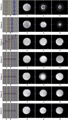
To examine the effects of changing current patterns, a ring with holder was designed (fig. 1b) (SolidWorks, Dassault Systèmes, Vélizy-Villacoublay, France) and 3D printed (RexRoi Europe, Leidschendam, The Netherlands). 9 patterns were designed and subsequently replicated on the ring, using copper wire. A major benefit of our design was that wire patterns could be changed and tested rapidly. Each pattern was evaluated and created the basis for the next iteration. All wire patterns are visible in figure 2.The crusher coil uses long wires that can cause RF-coupling, resulting in a distorted B1 field. To minimize this effect, high impedance elements, such as wire wound inductors are needed. At first, this was done by having meandering current wires. Later these were replaced by large loops and finally small loops.
After assembling each pattern, the ring was placed inside a 2 channel transmit - 32 channel receive coil (NOVA Medical). A bottle shaped oil phantom was placed in the middle of this ring. The setup was tested in a Philips 7T Achieva system. A 2D fast-field echo scan was used to evaluate the crushing efficiency for each setup. Scan parameters were as follows: FOV= 220x10x220, Voxel size= 5x5mm, Echo time= 3ms, Slice thickness= 10mm, TR= 300.000ms, TE= 3ms. The crushing field was generated using a 4 Ampere pulse from a 3rd order shim amplifier for 2ms after excitation and before signal readout.
Results & Discussion
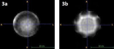
The meandering pattern of 1 and 2 resulted in uneven crushing (see fig. 3). This effect was particularly strong for pattern 2, which resulted in a star shaped crushing field. On the straight lines, there was strong crushing. It was concluded that the meandering will not create a homogeneous crushing field and a different approach to RF-decoupling was required.The next patterns were used to investigate the influence of straight lines. Figure 4 shows all remaining patterns, with two slices in the axial direction. Using pattern 3 and 4, clear image distortions appear which are removed when the small loops in the current direction are removed and replaced by loops in the Z-direction (visible from pattern 5 on).
Pattern 5 created hotspots located adjacent to the large loops. Pattern 6 compensated this effect with the use of a second return current leading to homogeneous crushing in the axial direction. The depth of the crushing however was reduced, likely due to a higher resistance of smaller loops. Crushing was improved by doubling the wires in pattern 7.
A coronal slide (Fig. 5a) of pattern 7 showed that the wire pattern does not create a homogeneous crushing field in coronal direction. This was attempted to be solved in patterns 8 (fig. 5b) and 9 (fig. 5c). Hotspots on the edges of the coil were ultimately reduced, but a homogeneous field was not created.
Conclusion
The tests that were performed gave insight in the current wire patterns of a crusher coil. The research actually showed that the meandering current lines cause severe inhomogeneities in the crusher field. Instead, it seems that optimal crushing in the axial direction is achieved through a pattern of concentric wires (pattern 6 to 9). Current wires that run in the direction of the B0 field should be negated by a current trace in the opposite direction. RF-coupling can be prevented by placing small loops in the Z-direction.The solution for homogeneous axial crushing can be used as a starting point for a design of current wires that create a homogeneous crushing field in the coronal direction. Future research will involve simulating the most promising wire patterns as a starting point for further optimization.
Acknowledgements
No acknowledgement found.References
1. Boer VO, van de Lindt T, Luijten PR, Klomp DW. Lipid suppression for brain MRI and MRSI by means of a dedicated crusher coil. Magnetic resonance in medicine : official journal of the Society of Magnetic Resonance in Medicine / Society of Magnetic Resonance in Medicine 2015;73(6):2062-2068. https://www.ncbi.nlm.nih.gov/pubmed/24947343
2. Huijing ER, Van Dijk L, Ali Haghnejad SA, Morsinkhof L, Klomp DWJ, Luijten PR, Wijnen JP, Bhogal AA. Next generation Crusher Coil for suppressing extra cranial lipid signals at 7 Tesla. ISMRM 2019, 0950.
3. Paul A. Yushkevich, Joseph Piven, Heather Cody Hazlett, Rachel Gimpel Smith, Sean Ho, James C. Gee, and Guido Gerig. User-guided 3D active contour segmentation of anatomical structures: Significantly improved efficiency and reliability. Neuroimage. 2006 Jul 1; 31(3):1116-28. [bibtex] [medline] [doi:10.1016/j.neuroimage.2006.01.015]
Figures


