4220
B0 shimming of the liver using a local shim coil array at ultra-high-field MRI1Radiology Department, UMC Utrecht, Utrecht, Netherlands, 2MR Shim GmbH, Reutlingen, Germany
Synopsis
Ultra-high-field MRI is more sensitive to variations in the B0 field specially for larger organs such as the liver. Dynamic shimming using a local array of shim coils can potentially improve the field homogeneity by 18-46% when compared to the standard hardware correction methods at 7T. The use of local shim coil arrays embedded in RF receiver coils may become a good strategy when maximizing shim performance considering the limited available number of shim channels, particularly for larger organs such as the liver at ultra- higher field strengths.
Introduction
Ultra-high field MRI (≥7T) has the potential to become a clinically viable tool due to its higher intrinsic SNR and CNR compared to lower magnetic field. This increase in SNR and CNR allows for higher spatial resolutions and higher sensitivity in image acquisition than conventional clinical MRI. However, one of the main technical challenges with increasing field strength is the static magnetic field inhomogeneity. Variations in the B0 field lead to local frequency offsets that can cause artefacts during MRI and MRS measurements.1 The magnetic susceptibility differences between tissue interfaces is one of the leading factors for local magnetic field inhomogeneities due to the linear increase in field offsets caused by tissue susceptibility at higher fields. To reduce the spatial variations, higher order spherical harmonics are needed in addition to the standard hardware B0 shimming methods of the scanner, which would require a high number of shim coils and channels.2 An alternative is the use of local shim coil arrays that can provide the high order of spatial field variation albeit confined to a small region. Moreover, local shim coils would couple less to the conductive bore thereby may have negligible eddy currents thus could generate spatially varying magnetic fields that counteract the B0 field variations in real time. Here, we demonstrate that B0 field variations in the liver can be substantially reduced using a local array of only 16 shim coils, when compared to the standard hardware correction method at 7T.Methods
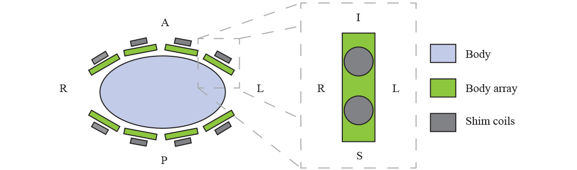
A whole-body 7T MR scanner equipped with a multi-transmit RF system (Achieva, Philips Health Care, Cleveland, OH, USA) was used to acquire B0 maps of the liver in five healthy volunteers. Eight transceiver fractionated dipole antennas with 16 additional receive loops interfaced to 8 parallel 2kW peak power amplifiers, were positioned symmetrically around the body at the position of the liver.3 RF phase shimming was performed to maximize transmission efficiency and optimize B1+ field homogeneity in the liver. Five dual-echo B0 maps (GE, 386×410×180mm3 FOV, 6×6×6mm3 voxel size, FA=4°, TR=6ms, TE=1.413ms, ΔTE=1ms) were acquired during free breathing as well as in inspiratory and expiratory breath-holds. The local shim coil array consisted of 16 circular loops, each with a diameter of 5cm. The coil diameter was optimized using simulation libraries provided by MR Shim GmbH (Reutlingen, Germany) between the range of 1-10cm. Each loop consisted of 20 coil windings of enameled copper wire with a diameter of 0.56mm.4 After the diameter was optimized, electromagnetic simulations were performed using 30 slices of the gradient dual-echo field maps to calculate the magnetic field that is generated by a constant electric current through the shim coil array using the Biot-Savart Law.5 The coils were arranged in two rows of eight around the body (Figure 1). These simulations were validated afterwards with a measurement in the 7T MRI scanner. B0 maps with equal sequence parameters were acquired in a phantom (octagon, 39×40×20cm, filled with polyvinylpyrrolidone) using a single shim coil driven with a current of 8A. The difference between the simulation and the measured B0 map was calculated as the RMSE and the correlation with the Pearson correlation coefficient.6Results
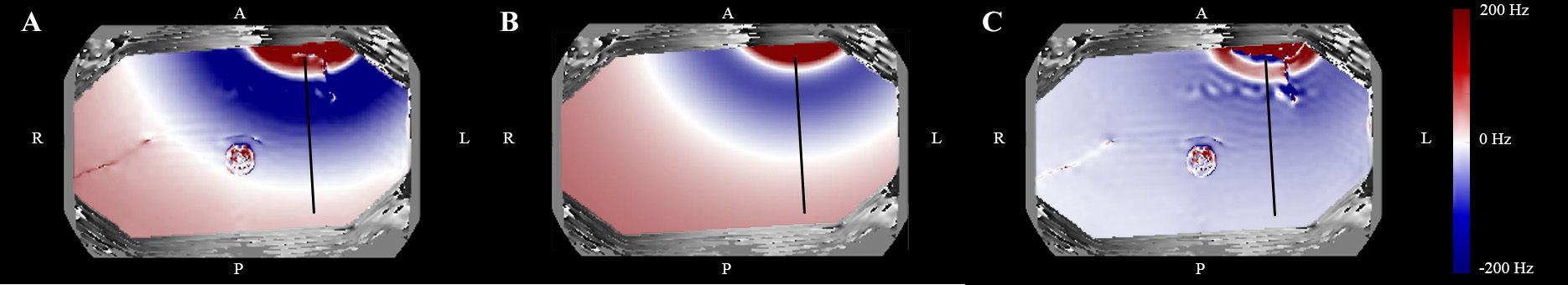
The simulations showed that the use of the local array of shim coils led to 65±8.3% improvement of the B0 homogeneity during free breathing, 80±4.5% improvement during inspiratory state, and 75±12% improvement during expiratory state when compared to unshimmed B0 map acquired during free breathing (Figure 2, 3). Compared to the field homogeneity when using the standard hardware-based method up to second order terms, the B0 variation in the liver can be reduced by 9±5.1% in free breathing, 46%±16 in inspiratory state, and 18±12% in expiratory state. Comparison of the simulations with the phantom 7T measurements, shows clear B0 differences (Figure 4, 5). The difference is close to zero further away from the shim coil. The Pearson correlation coefficient between simulations and measurements is 0.70 and the RMSE is 196Hz with the maximum B0 field variation value measured at 5.34×10³Hz and the maximum of the simulation at 1.66×10³Hz.Discussion
Substantial gain in field uniformity can be obtained with organ specific local B0 shim coils when compared to generalized static spherical harmonic shimming. In our study, we simulated dynamic shimming by using breath-hold, while real-time shim updating was not implemented. Direct use of simulated fields in real time will cause a mismatch as shown by the phantom measurements that differed from the simulations. While this can be prevented by obtaining fast B0 calibration maps from all shim coils, the magnitude of field variation is similar, thereby the simulation results do provide validation for the power requirements of the coils and amplifiers.Conclusion
This study shows that local B0 field variations in the liver at 7T can be reduced by using an array of local shim coils. B0 shimming improves by 9±5.1% in free breathing up to 46% in inspiration when using a local shim coil array in comparison to standard hardware shimming B0 shimming. As local shimming becomes more important at ultra-high magnetic field strengths, the use of embedding local shim coils in RF receiver arrays may become attractive, particularly for larger organs such as the liver.Acknowledgements
No acknowledgement found.References
1. Truong Trong-Kha, et al. Effects of static and radiofrequency magnetic field inhomogeneity in ultra-high field magnetic resonance imaging. Magnetic Resonance Imaging. 2006; 24:103-112.
2. Pan JW, Lo KM, Hetherington HP. Role of high order and degree B0 shimming for spectroscopic imaging of the human brain at 7 Tesla. Magn Reson Med 2012; 68:1007-1017.
3. Raaijmakers AJE, Italiaander M, Voogt IJ, Luijten PR, Hoogduin JM, Klomp DWJ, van den Berg CAT (2015) The fractionated dipole antenna: a new antenna for body imaging at 7 Tesla. Magn Reson Med 75:1366–1374.
4. MR Shim GmbH, Reutelingen, Germany
5. Juchem C, Nixon TW, McIntyre S, Boer VO, Rothman DL, de Graaf RA. Dynamic multi-coil shimming of the human brain at 7 T. Journal of Magnetic Resonance 2011; 212(2):280-288.
6. Fisher, R.A. Statistical Methods for Research Workers, 13th Ed., Hafner, 1958.
Figures