4206
Study of gradient induced electric fields in a tissue-mimicking ASTM phantom within an X-Axis gradient coil
Arjama Halder1,2, Ali Attaran2, William B Handler2, and Blaine A Chronik2
1Medical Biophysics, Western University, London, ON, Canada, 2Physics and Astronomy, Western University, London, ON, Canada
1Medical Biophysics, Western University, London, ON, Canada, 2Physics and Astronomy, Western University, London, ON, Canada
Synopsis
Validating measured gradient induced electric fields in a conductive ASTM phantom within an X-Axis gradient coil.
INTRODUCTION
The objective of this study was to investigate gradient coil induced electric fields using an ultra-low frequency electric field probe within a conductive ASTM phantom in an operating gradient coil. In recent years there has been a growing population of people equipped with active implantable medical devices (AIMDs) who might require an MRI exam at some point in the future. However, the electromagnetic interactions between devices and the various sub systems of MR scanners lead to potential hazards that can cause patient to have limited, to no access to an MRI1. The ISO/TS 10974:20182 mentions one such potential cause of worry, to be resulting from the electromagnetic interaction between the MR scanner pulsed gradient fields and AIMDs. Electric field quantification is one of the most difficult aspect of this interaction and can cause patient harm of varying severity, depending on the AIMD type.Most of the studies performed to address device compatibility with the MR scanner that focuses on this issue concentrates on designing different test platforms that mimics the dB/dt fields produced by gradients2,3 or developing test methods that allow electric field measurements without the need of generating dB/dt fields4. Here we propose the use of an ultra-low frequency electric field probe5,6, as a supplementary test method to measure the actual variation of the electric field caused by an operating gradient coil in a scanner. The use of this probe is validated using a saline filled ASTM phantom as a representation of a tissue-mimicking environment and a high performance asymmetric head gradient coil outside of the scanner. Performing this test with sufficient accuracy in an operating gradient coil is difficult given the strength of the field generated, but can be very useful for direct field measurements in the scanner for device testing purposes, and also validates electric fields predicted using the low frequency solver in Sim4Life (Zurich Med Tech).
METHOD
To perform these measurements the ASTM phantom was filled with saline up to the height of 9 cm (conductivity of saline = 0.47 S/m) and placed within a gradient coil as shown in figure 1. For this set of measurement, only the X-Axis of the gradient coil was used, with the a sinusoidal current of 126 A at a frequency of 1 kHz. This operation frequency allowed the use of quasi-static approximations7 which led us to assume that tissues are uniform volume conductors. The ultra-low frequency electric field probe6 was held and moved by a robotic arm which allowed precise measurements at locations near the head region of the phantom. The probe was constructed using a 2 cm short dipole antenna followed by a 2-stage instrumentation amplifier and a differential amplifier that provided differential amplification and removed common-mode voltage respectively. This probe had a differential input impedance of 0.8||2 (GΩ||pF), while the designed amplifier had a gain of 100 and a cut-off frequency of 31 kHz. Data was collected via a connection to an oscilloscope with a termination impedance of 1 MΩ using an RG58 cable. To remove the extrinsic influences in the measurements the saline solution was grounded.Post-processing of the collected data involved removing high frequency noise from the measurements using a low-pass filter for every position. A sinusoidal fit was performed on the filtered signals to obtain the peak values at those positions. To quantify the error of the fit a root mean squared error (RMSE) was calculated for each fit performed. The fitted signal was then scaled with the calibration factor associated with the probe, which is 2.64 (V/m)/V. Simulations were performed in Sim4Life using the magneto-quasi-static low frequency solver to compare against the experimental results.
RESULTS
The experimental setup along with a schematic is shown in figure 1. Figure 2 shows the simulated electric field magnitude on the xz, yz and xy plane at the center of the phantom. Figure 3 show the measured and simulated results along a line y = 0 on 2 different yz plane that are about 3 cm off in either direction from the center of the phantom with the probe oriented in the z direction.DISCUSSION
The experimental results show a reasonable agreement with the simulated behavior of the electric field given the sensitivity of the field measured, as shown in figure 3. This agreement shows that this prototype electric field probe makes it possible to obtain useful measurements of electric fields induced by gradients inside tissue. Future measurements will build upon this to explore the full extent of the phantom and the other gradient axes. Measurements will also be performed with an AIMD within the phantom. The results achieved from the this study show possible commercial use of such a probe for conducting gradient induced electric field measurements in AIMDs within a scanner or a test platform.CONCLUSIONS
ISO/TS 10974:20182 considers gradient induced extrinsic electric fields to a potential cause of worry for patients with AIMDs. The standard proposes radiated gradient immunity test to account for these effects. This research provides a supplementary test method that allows direct measurement of the variation in electric field using an ultra-low frequency electric field probe within a gradient coil.Acknowledgements
The authors would like to thank the research and financial supports received from Natural Sciences and Engineering Research Council (NSERC) of Canada, the Ontario Research Fund (ORF), and CMC Microsystems.References
- Gimbel JR, Kanal E. Can patients with implanted pacemakers safely undergo magnetic resonance imaging? J. Amer. College Cardiol.2004;43(7):1325–1327.
- International Organization for Standarization. Assessment of the safety of magnetic resonance imaging for patients with an active implantable medical device. ISO/TS 10974. 2018;4.
- Martire DJ, Handler WB, Chronik BA. Design of an MRI Gradient Field Exposure System for Medical Device Testing. ISMRM. 2017; Abstract # 4334.
- Xin X, Chen XL, Sison S. A MRI Gradient Induced Electric Field Exposure System for Active Implanted Medical Devices. ISMRM. 2019; Abstract # 1440.
- Glover PM, Bowtell R. Measurement of Electric Fields Due to Time-Varying Magnetic Field Gradients Using Dipole Probes. Physics in Medicine and Biology. 2007;52: 5119-5130.
- Halder Arjama Attaran A, Handler WB, Chronik BA. Electric Field Probe used for Gradient Coil Induced Field Measurements During Medical Device Testing: Design, Calibration and Validation. IEEE Transactions on Antennas and Propagation. 2019; ID # AP1907-1282.R1. (Under review)
- Roth BJ, Cohen LG, Hallett M. The Electric Field Induced During Magnetic Stimulation. Magn Mot Stimul Basic Princ Clin Exp. 1991;43:268-278.
Figures
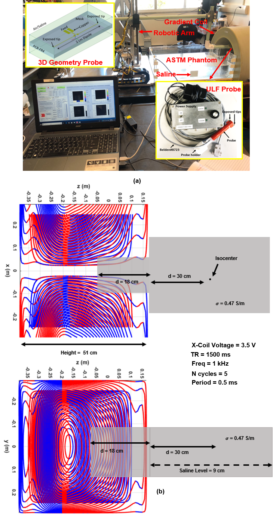
Fig. 1. (a) The experimental
setup and the design of the ultra-low frequency electric field probe. (b) The
schematic for the setup along with the information of the X-Axis gradient
operation.
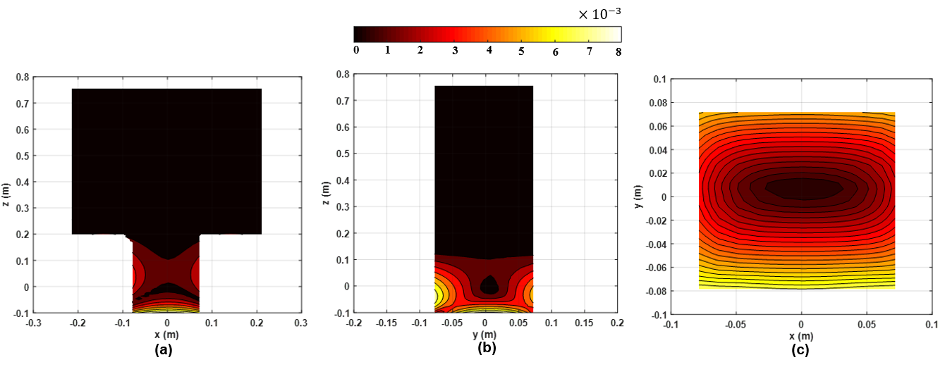
Fig.2. The simulated
electric field magnitude on the (a) xz plane (b) yz plane and (c) xy plane at
the center of the phantom.The peak value for current for this simulation was
set to 1 Amps.
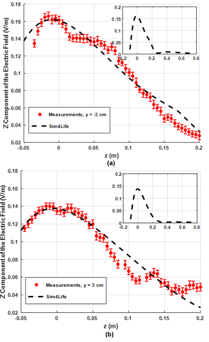
Fig. 3. The main plot compares the measured with the simulated
electric field along a line y = 0 on 2 different
yz plane with the probe orientated in the z direction (a) The first yz plane is
located 3 cm away in the negative direction from the center of the phantom. (b)
The second yz plane is located 3 cm away in the position direction from the
center of the phantom. The subplots in both cases show the overall simulated behaviour of
the field as a function of location. The peak value for current during this
experiment was set to 126 A.