4192
Computational Study of RF Induced Heating and Safety for Tattoo Patients under MRI1Biomedical Engineering, Hanyang University, Seoul, Republic of Korea, 2Center for Research Equipment, Korea Basic Science Institute, Daejeon, Republic of Korea
Synopsis
Patients with permanent cosmetics and tattoos have potential risk of heating effects during MRI examination. Most of the cutaneous reaction and injury occurred around the tattoos due to the metallic pigments found in the tattoo inks. Previous reports and studies presented that mild edema and redness of the tattoo comprehended as cutaneous burn due to the shape of the tattoo. This study numerically analyzed the RF heating effects around the tattoo with different types and positions. Results show that high SAR and temperature rise were observed around the circular loops, long strips and adjacent point of the tattoo.
Purpose
When a patient with permanent tattoos and cosmetics performs the MRI examination, there is a potential risk of RF induced heating and power deposition around the tattoos due to time varying magnetic field (B1) of the RF birdcage coil. A significant challenge concerns the precise characterization of tattoo material, shapes and types that might produce a burning sensation of the skin around the tattoos during MRI examination. Several types of tattoo pigments are sensitive to magnetic field, which can cause the adverse effects in the clinical events, such effects may increase the temperature of the tattoo pigments [1] - [2]. A question arises here, whether individual color pigments or class of tattoo pigments with physical behavior or specific characteristic cause burning sensation. Moreover, the risk of burning is especially high when the metallic pigment (iron oxide) is organized in the form of sharp edges, multiple adjacent points and large circular loops of the tattoos [3]. The previous reports are lacking the detailed studies of the tattoo effects under clinical MRI examination. The purpose of this study to evaluate the tattoo effects in term of temperature rise and RF energy deposition at different field strength of the MRI.Methods
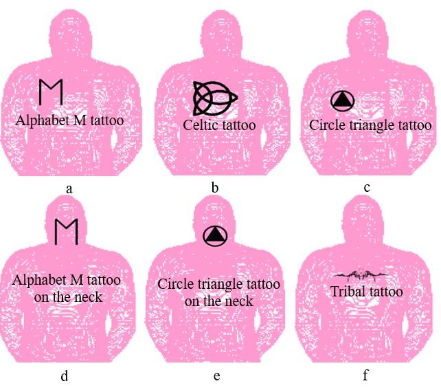
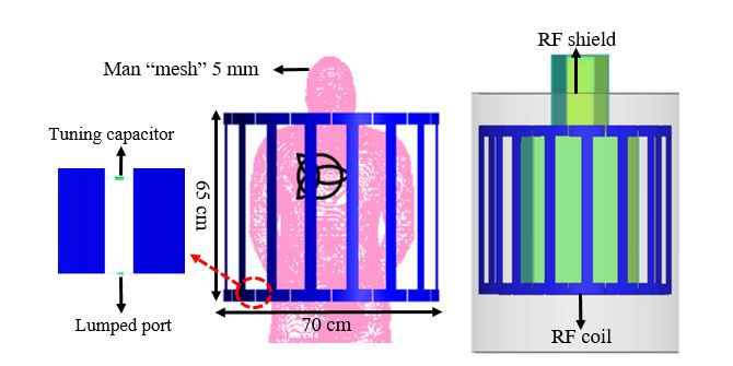
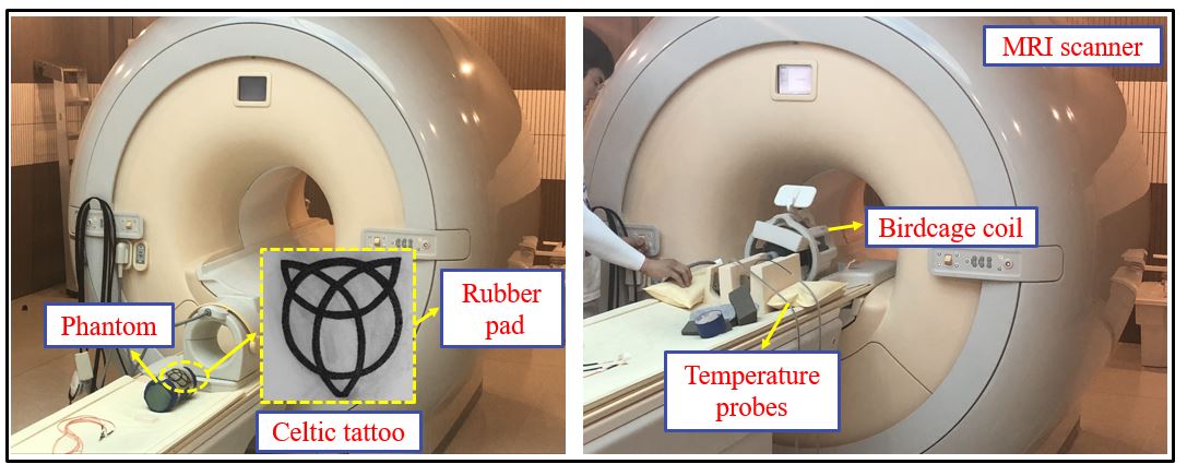
To compare the difference of field distribution between patients with different types and positions of tattoo, six types of condition were modeled as depicted in Fig. 1. Fig. 2 shows the high pass birdcage coil, which were tuned at the different field strength of MRI by changing the capacitor values. Four different types of tattoo and two different positions were chosen to investigate the RF induced heating and temperature rise around the tattoos. It should be noted that the typical tattoo have complex structures, and reproduced the exact tattoos via simulations are not possible; however, an approximate model of the tattoo can be made. Bio heat and finite-difference time-domain (FDTD) based electromagnetic simulations are carried out to study the temperature rise and specific absorption rate (SAR) distributions [4]. Fig. 3 present the experimental setup of 3 T MRI with a celtic tattoo. The celtic tattoo is designed on the rubber pad and placed on the cylindrical phantom, which is filled with Gel-saline solutions. This study presents a case study of tattoo with different shapes and positions on the realistic human model to investigate the heating effects.Results
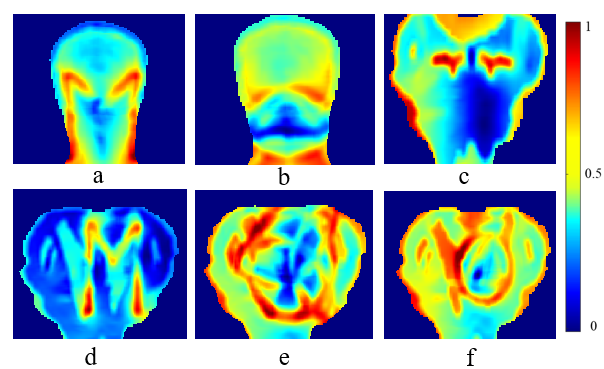
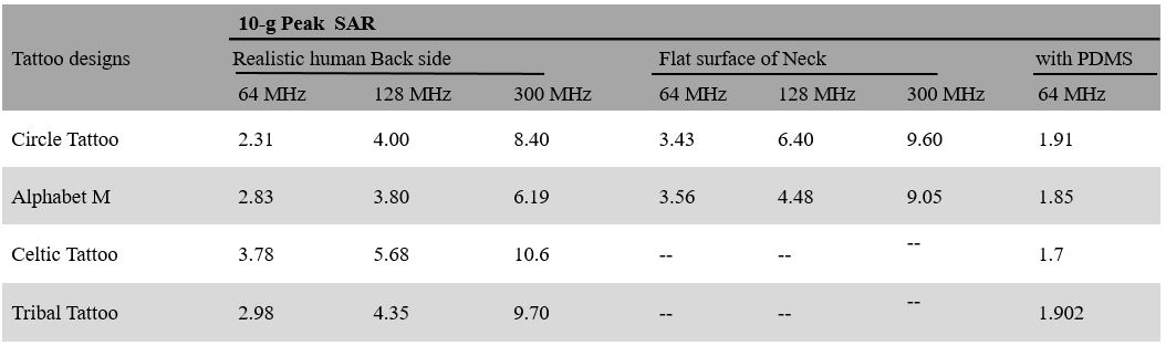
Simulation results show that the maximum local SAR are positively correlated with tattoo shapes. The hot spot for the tattoos were observed on the long strip, sharp edges, and multiple adjacent points of the tattoo. Based on the types of the tattoo, we analyzed that how the RF induced heating depends on the shapes of the tattoo. The distribution of 10-g average SAR of the tattoos for neck and back of the human phantom are depicted in Fig. 4. When metallic tattoos are immersed in the RF field of MRI, the incident field is coupled and scattered at the adjacent points, strips and edges of the tattoos. Because of the mismatching impedance between the tattoos and tissues of the skin, extra amounts of scattered electric field occur at the different position of the tattoos. Due to ohmic loss of human tissues, the energy is converted into heat, which cause a cutaneous reaction to the skin. Furthermore, polydimethylsiloxane (PDMS) plays an important role in biological and chemical applications, due to its biocompatibility, mechanical compliance, optical transparency, ease of fabrication, and chemical stability [5]. The results present that the PDMS is effective to reduce SAR distribution around the tattoos. It can be observed from the Fig. 5 that the PDMS reduced SAR in the region of interest at 1.5 T. All the simulated results were normalized to the whole body average SAR of 2 W/kg. The table I present peak 10-g average SAR due to the shape of tattoos. Based on the table I results, we investigated that the SAR value for the celtic tattoo is almost double compared to the circle triangle tattoo due to the sharp edges and large circular loops at 1.5 T. These results show the interaction between RF field of MRI and tattoo depends on the shapes and types of the tattoo.Conclusion
In this study, the effect of tattoos at different field strength of MRI was evaluated. This study present that the shape of the tattoo (i.e. large circular loops, sharp edges and multiple adjacent points) is the major factor to increase the RF induced heating. The procedures presented in the simulation environment are used to facilitate the RF safety assessments for tattoo patients at both ultra-high field strengths and clinical MRI.Acknowledgements
This work was supported by the Basic Science Research Program through the National Research Foundation of Korea funded by the Ministry of Science and ICT under Grant 2019R1A2C2004774.
References
- Kluger N, Brun‐Lévêque P, Gral N. Painful burning sensation on a tattoo during magnetic resonance imaging. International journal of dermatology. 2019 Apr;58(4):E82-3.
- Vahlensieck M. Tattoo-related cutaneous inflammation (burn grade I) in a mid-field MR scanner. Eur Radiol. 2000;10(1):197–197.
- Ross JR, Matava MJ. Tattoo-induced skin “burn” during magnetic resonance imaging in a professional football player: a case report. Sports Health. 2011;3(5):431-434.
- Sim4life by ZMT, https://www.zmt.swiss.
- Du P, Lin X, Zhang X. Characterization on the Electrical Properties of PDMS Nanocomposites by Conducting Polymer Nanowires. MRS Online Proc Libr Arch. 2011;1312.
Figures