4182
MRI safety of deep brain stimulation devices: Radiofrequency heating of a commercial lead and an insulated copper wire1Physical Sciences, Sunnybrook Research Institute, Toronto, ON, Canada, 2Electrical and Computer Engineering, McMaster University, Hamilton, ON, Canada, 3Division of Neurosurgery, Sunnybrook Health Sciences Centre, Toronto, ON, Canada, 4Harquail Centre for Neuromodulation, Sunnybrook Research Institute, Toronto, ON, Canada, 5Medical Biophysics, University of Toronto, Toronto, ON, Canada
Synopsis
Magnetic resonance imaging (MRI) safety of deep brain stimulation (DBS) patients remains a concern. Because of this, researchers have relied on phantoms to mimic electromagnetic behavior. DBS devices are costly and as a result, copper wire is often used as a substitute to DBS leads. This work examines the suitability of using copper wire to emulate MRI-DBS interactions with results showing over 50 % reduction in temperature elevations when using a DBS lead.
Introduction
Usage of deep brain stimulation (DBS) treatment continues to grow worldwide. Magnetic resonance imaging (MRI) can play an important role in the workflow to fit patients with DBS devices, but there are safety concerns that must be considered when imaging is conducted post-implantation. In these situations, the radiofrequency (RF) electromagnetic field can induce electrical current to flow along the device, increasing the possibility of exceeding the present MRI safety guidelines for the local specific-absorption rate (SAR) and in more severe incidences, causing tissue damage. Researchers thus far have relied heavily on phantom studies for safety investigations in advance of patient imaging, and for demonstrating new methods to enhance MRI safety in proof-of-concept1,2. Often, simple homogenous phantoms and/or copper wires are used respectively to emulate the electromagnetic behaviour of human tissue and commercial DBS leads during MRI. Given the simple construction of such setups, however, the level of realism can be somewhat limited. Alternatively, complex heterogeneous anthropomorphic phantom structures can be designed and assembled quite easily using 3-D printing methods, with inclusion of actual DBS devices. Cost becomes more of a factor in this case, as DBS devices are expensive. These considerations make it important to examine the suitability of using copper wires to emulate MRI-DBS conditions and to our knowledge a direct comparison has not been investigated before. The present work addresses this aim by investigating the use of copper wire versus a commercial DBS lead in two RF heating experimental setups.Methods
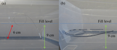
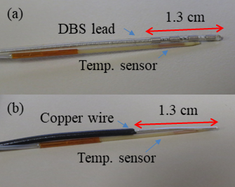
All RF heating experiments were conducted on a 3 T MRI system (Magnetom Prisma, Siemens) configured for body coil RF transmission and reception using a turbo spin-echo (TSE) pulse sequence (TR / TE = 6000 ms / 103 ms, flip angle = 165, duration = 1:38 mins). A 19 cm x 33 cm x 13 cm rectangular container was filled with a poly-acrylic acid (10 g/L) and salt (1.32 g/L) solution that produced a conductivity of ~0.47 S/m and relative permittivity of ~80. The gel-based solution filled the container to a depth of 9 cm. A 40-cm long commercial DBS lead (3387, Medtronics, Minneapolis, MN) and a 40-cm long insulated copper wire (AWG 22) with a 1.3 cm exposed tip were used in two experimental setups: 1) where the DBS lead and copper wire were routed such that the looped section (or majority thereof) was externalized from the phantom solution with the tip-end submerged to an approximate depth of 6 cm into the phantom solution, as shown in Fig. 1a using the DBS lead; and 2) where the DBS lead and copper wire were fully immersed into the phantom, as shown in Fig. 1b using the insulated copper wire. Temperature measurements for RF heating effects were recorded at locations near the lead and wire tips as shown in Fig. 2 using fibre-optic temperature sensors (Opsens Inc., Quebec City, QC). Control measurements were recorded at the opposite end of the container using a fibre-optic temperature sensor (Neoptix, Québec City, QB) to verify that RF heating effects were localized.Results
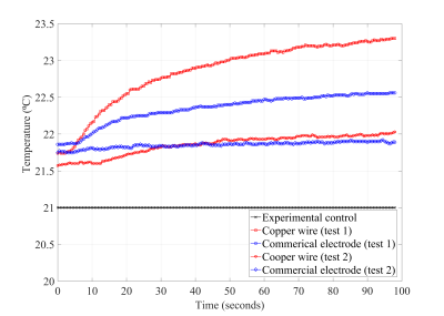
Fig. 3 plots the temperature elevations for TSE imaging for all test scenarios. Temperature increases for setup 1 were 1.6 ± 0.3⁰C when imaging with the copper wire and 0.7 ± 0.3⁰C when imaging with the DBS lead. For setup 2, temperature increases were 0.4 ± 0.3⁰C for the copper wire and 0.1 ± 0.3⁰C for the DBS lead. Fig. 4 shows a sample TSE image at a slice intersecting the DBS lead and copper wire.Discussion and Conclusion
The experimental results showed that the commercial DBS lead was more resistant to RF electromagnetic coupling than the conventional copper wire. The temperature increase obtained from the DBS lead was over 50 % lower in both setups. Engineering design is likely the key contributor to this difference. For example, the DBS lead is a multi-layer structure that uses platinum-iridium wire and fluoropolymers for insulation. The overall result suggests moving toward a complex phantom structure with a commercial DBS device is advisable for improved realism, while highlighting the significant difference in temperatures measured, a useful observation for researchers in the field to consider.Acknowledgements
No acknowledgement found.References
[1] Kahan et al., “The Safety of Using Body-Transmit MRI in Patients with Implanted Deep Brain Stimulation Devices.” PLOS One 2015.
[2] McElcheran et al., “Investigation of Parallel Radiofrequency Transmission for the Reduction of Heating in Long conductive Leads in 3 Tesla Magnetic Resonance Imaging. PLOS One 2015.
Figures


