4173
Magnetic Resonance Imaging-guided Focused Ultrasound ablation of lumbar facet joints of a patient with a MRI non-conditional pacemaker at 1.5T1Radiology, Mayo Clinic, Rochester, MN, United States
Synopsis
An MRgFUS ablation treatment of lumbar facet joints in a patient with a traditional MRI non-conditional pacemaker was completed. A risk-benefit analysis by a coordinated multi-disciplinary team prior to this treatment was performed to account for the risks associated with traditional MRI non-conditional pacemaker. The treatment was successfully performed as per our institution established cardiac implanted electronic device (CIED) MRI practice and the patient had no adverse cardiac event during or following this procedure. By careful use of our institutional CIED MR-practice guidelines, we demonstrated that such treatments can be safely achieved for patients with CIEDs on a case-by-case basis.
INTRODUCTION
Magnetic Resonance Imaging-guided Focused Ultrasound (MRgFUS) is a minimally-invasive treatment modality that utilizes an ultrasound transducer integrated in an MRI scanner. MRgFUS systems have been successfully used to treat symptomatic uterine fibroids 1-4 and facet joint pain 5. During MRgFUS treatment, ultrasound energy is focused within target tissues causing localized thermal ablation; MRI is essential for treatment planning, US beam guidance, real-time MR-thermometry and treatment assessment 3. Facet joint treatments in our practice utilize an ExAblate 2100 MRgFUS system (Insightec, Haifa, Israel) integrated with a 1.5T MR scanner (Signa Excite, General Electric, Waukesha , WI), which poses significant safety challenges for patients with implanted cardiac devices potentially excluding them from treatment. This study describes a MRgFUS ablation of lumbar facet joints in a patient with a traditional MRI non-conditional pacemaker, there has been a dearth of such treatments reported 6.METHODS
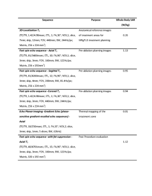
In this study an 80 years male with a history of chronic axial lower back pain underwent MRgFUS ablation of the bilateral L3-4, L4-5, and L5-S1 facet joints. The patient had a history of symptomatic sinus bradycardia which was managed with a dual chamber transvenous pacemaker (Assurity DR2240, MRI non-conditional, St Jude Medical, Memphis, TN); the patient was not pacemaker-dependent. A risk-benefit analysis was carried out prior to this ablation treatment to account for the risks associated with MRI non-conditional pacemaker, and the exam was performed in accordance with our established cardiac implanted electronic device (CIED) / MRI-practice, with over 3000 patients scanned, that includes a coordinated team of physicians, cardiology pacing nurses, MRI physicists and technologists. Prior to entry into MR scanner room the cardiology personnel programmed the patient’s pacemaker to DOO (dual-absence of sensing - no response to sensed input) mode of 80bpm. The initial transducer localization within MRI coordinates for the ablation treatment required acquisition calibration scans based on single-shot fast spin echo pulse sequence. For this patient the 1.5W/kg limit was exceeded in the scanner-based SAR estimation for the corresponding patient weight of 77kg where the SAR was predicted to be 1.73W/kg. Therefore, this calibration scan was completed using a QA phantom. The patient was subsequently positioned into the feet first-supine position on top of the MRgFUS table with his lower back located directly above the transducer. After this process the table was slowly advanced into the iso-center of the MR bore. For the entirety of the treatment the cardiology nurse continually monitored the patient’s cardiac function through electrocardiography. The MRI was performed in normal operating mode for both SAR and gradient switch rates. All MR sequences executed during the 206mins of the procedure were adjusted to maintain the whole-body SAR below the threshold of 1.5W/kg, which is a consensus value for safe scanning of patients with non-MR conditional pacemakers at our institution 7. Furthermore, the real-time SAR display was monitored and sequences were stopped when the 10s SAR average exceeded the 1.5W/kg threshold.RESULTS
Following a 3D scout (0.26W/kg), a set of three anatomical T2-weighted fast spin echo sequences, in axial, sagittal, and coronal planes, were acquired (SAR =1.13, 0.95, and 0.94W/kg) for treatment planning (Table 1). These images were then transferred to the ExAblate workstation where the target locations of the MRgFUS ablation were planned using the ExAblate treatment planning system. MRgFUS of the bilateral selected lumbar facet joints was performed using 22 individual sonications 5. Each sonication was monitored in the axial plane using phase-sensitive gradient-recalled echo sequences acquired for the purpose of MR thermometry feedback with temporal resolution of 6s. The images were transferred in real-time to the ExAblate workstation where the thermometry and the corresponding treatment dose maps were overlaid onto the patient anatomy. Upon completion of the treatment T2-weighted fast spin echo sequence with fat suppression (1.12W/kg) was acquired to assess for edema around the target joints. The patient had no adverse cardiac event during or immediately following the MRgFUS procedure. His pacemaker was subsequently interrogated, which confirmed no damage or alteration, and it was reprogrammed to its original settings. Informed consent for publication purposes was obtained from the patient.DISCUSION
MRgFUS ablation of the facet joint is a new treatment being used for facet joint pain 5, 8, 9 which is dependent upon the ability to accurately image the area undergoing treatment in real-time using MRI. A large proportion of the patient group with facet joint pain tend to be an older population with confounding cardiac morbidities often with a CIED 10. Despite the fact that these devices are non-MR conditional are regarded as presenting contraindications to MRI 11, 12, our institution has under certain precautions safely scanned over 3000 such devices over the last ten years 7. By adopting and incorporating these guidelines into the process of an MRgFUS procedure it was possible to safely complete a facet joint ablation with no changes in pacemaker function or pacing threshold, or any other complications.CONCLUSION
This study reports on successful MRgFUS lumbar facet joint ablation in a patient with a MRI non-conditional pacemaker. By careful use of our MRI CIED protocol 7, we demonstrated that the MRgFUS ablation treatment of facet joints can be safely achieved for patients with CIEDs on a case-by-case basis.Acknowledgements
NoneReferences
1.Gorny KR, Woodrum DA, Brown DL, Henrichsen TL, Weaver AL, Amrami KK, et al. Magnetic resonance–guided focused ultrasound of uterine leiomyomas: review of a 12-month outcome of 130 clinical patients. Journal of Vascular and Interventional Radiology. 2011;22(6):857-64.
2.Hesley GK, Felmlee JP, Gebhart JB, Dunagan KT, Gorny KR, Kesler JB, et al., editors. Noninvasive treatment of uterine fibroids: early Mayo Clinic experience with magnetic resonance imaging-guided focused ultrasound. Mayo Clinic Proceedings; 2006: Elsevier.
3.Hesley GK, Gorny KR, Henrichsen TL, Woodrum DA, Brown DL. A clinical review of focused ultrasound ablation with magnetic resonance guidance: an option for treating uterine fibroids. Ultrasound quarterly. 2008;24(2):131-9.
4.Hesley GK, Gorny KR, Woodrum DA. MR-guided focused ultrasound for the treatment of uterine fibroids. Cardiovascular and interventional radiology. 2013;36(1):5-13.
5.Tiegs-Heiden Christin A LVT, Gorny Krzysztof R. , Boon Andrea J. , Hesley Gina K. . A Case of Improved Treatment Response Following MR-guided Focused Ultrasound for Lumbar Facet Joint Pain. Mayo Proceedings. 2019;In press.
6.Ito H, Fukutake S, Yamamoto K, Tanaka S, Yamaguchi T, Taira T, et al. Magnetic Resonance Imaging‐guided Focused Ultrasound Thalamotomy for Parkinson's Disease with Cardiac Pacemaker: A Case Report. Movement disorders clinical practice. 2018;5(3):339.
7.Padmanabhan D, Kella DK, Deshmukh AJ, Mulpuru SK, Mehta RA, Dalzell CM, et al. Safety of Thoracic Magnetic Resonance Imaging for Patients with Pacemakers and Defibrillators. Heart rhythm. 2019.
8.Harnof S, Zibly Z, Shay L, Dogadkin O, Hanannel A, Inbar Y, et al. Magnetic resonance-guided focused ultrasound treatment of facet joint pain: summary of preclinical phase. Journal of Therapeutic Ultrasound. 2014;2(1):9.
9.Krug R, Do L, Rieke V, Wilson MW, Saeed M. Evaluation of MRI protocols for the assessment of lumbar facet joints after MR-guided focused ultrasound treatment. Journal of Therapeutic Ultrasound. 2016;4(1):14.
10.Grewal SS, Gorny KR, Favazza CP, Watson RE, Kaufmann TJ, Van Gompel JJ. Safety of Laser Interstitial Thermal Therapy in Patients With Pacemakers. Operative Neurosurgery. 2018;15(5):E69-E72.
11.Strom JB, Whelan JB, Shen C, Zheng SQ, Mortele KJ, Kramer DB. Safety and utility of magnetic resonance imaging in patients with cardiac implantable electronic devices. Heart rhythm. 2017;14(8):1138-44.
12.Sommer T, Luechinger R, Barkhausen J, Gutberlet M, Quick H, Fischbach K, editors. German Roentgen Society Statement on MR imaging of patients with cardiac pacemakers. RöFo-Fortschritte auf dem Gebiet der Röntgenstrahlen und der bildgebenden Verfahren; 2015: © Georg Thieme Verlag KG.
