4166
Concurrent alternating transcranial current stimulation and fMRI: subject safety and the data quality evaluation
Beni Mulyana1,2, Qingfei Luo1, Aki Tsuchiyagaito1, Ashkan Rashedi1, Jared Smith1, Masaya Misaki1, Duke Shereen3, Samuel Cheng2, Martin Paulus1, Hamed Ekhtiari1, and Jerzy Bodurka1,4
1Laureate Institute for Brain Research, Tulsa, OK, United States, 2University of Oklahoma, Tulsa, OK, United States, 3The Graduate Center, City University of New York, New York, NY, United States, 4Stephenson School of Biomedical Engineering, University of Oklahoma, Norman, OK, United States
1Laureate Institute for Brain Research, Tulsa, OK, United States, 2University of Oklahoma, Tulsa, OK, United States, 3The Graduate Center, City University of New York, New York, NY, United States, 4Stephenson School of Biomedical Engineering, University of Oklahoma, Norman, OK, United States
Synopsis
We evaluated subject safety and the fMRI image quality in the presence of transcranial alternating current stimulation (tACS). Scalp temperatures under the tACS electrodes with and without stimulations were measured. Result indicated: i) the tACS stimulation itself had no harmful impact on scalp temperature; ii) tACS’s artifacts had negligible effects on fMRI images. Although the fMRI scan produced heating effects due to the RF energy deposition in tACS electrodes, the scalp temperatures remained below 37°C during a 12-min scan regardless of the presence of stimulations. Thus, concurrent tACS with fMRI has no impact on patient safety and image quality.
INTRODUCTION
Increasing evidences showed that the transcranial alternating current stimulation (tACS) could modulate brain activities, and combining tACS and functional MRI (tACS-fMRI) are considered to a promising way to reveal the causal relationships between brain stimulation and brain activity [1-4]. Before applying tACS-fMRI, it is necessary to verify whether tACS-fMRI has impact on subject safety and fMRI image quality. The study aims were: (1) to conduct tACS safety test in the aspect of the scalp temperature during the stimulation, and (2) to examine whether tACS stimulation would induce any artifacts on fMRI images.METHOD
Experiments were performed using a 3T MRI scanner (Discovery MR750; GE Healthcare Systems, Milwaukee, WI) with the 8-channel receive-only head coil. One healthy participant experienced three gradient-echo EPI scans with tACS (Starstim R32; Neuroelectrics Barcelona SLU; Spain) stimulations. The image acquisition parameters were: FOV = 240 × 240 mm, matrix = 96 × 96, 39 axial slices with slice thickness = 2.9 mm. In the 1st run, EPI images were acquired without the RF excitation to evaluate the impact of tACS on the thermal noise in images. The tACS montage5 was set with F3 and F4 as stimulations (1mA, 6Hz, 0 phase, 8cm2 pad), and Cz as a return (8cm2 pad). We adopted the block design with 2-min/2-min stimulation ON/OFF during the 6-min EPI scan. The mean and standard deviation (SD) of images during stimulation ON and OFF were compared to evaluate the effects of the tACS stimulation on the EPI noise.Next, we obtained two scanning runs to conduct safety tests by placing temperature sensor (Biopac TSD202A) on the scalp under the electrodes. Prior to the scanning run, we measured the baseline temperatures on the scalp under the tACS electrodes without any scan nor tACS stimulations. After measuring the baseline temperatures, we obtained one 12 mins EPI run (2nd run) (with RF excitation) leaving the same tACS parameters as the first run to investigate whether there are no heating effects of tACS stimulation on the scalp. We repeated one more EPI run (3rd run) with a bilateral tACS montage, in which electrodes F4 and P4 were used as stimulations and Cz as a return.
RESULTS
Figure 1 shows the result of the noise test (1st run). The average and SD of image noise from spherical voxels (radius=1cm) under F4 and F3 were not significantly different between tACS stimulation ON and OFF (ON: mean = 61.61, SD = 13.89; OFF: mean = 58.76, SD = 20.42, t(59) = 0.95, p = 0.34). In addition, no tACS-related artifacts were observed in the EPI images (Fig. 2). Figures 3 and 4 show the results of the tACS safety test. The baseline scalp temperatures prior to scanning and tACS stimulation were stable around 33°C (Fig.3). The EPI scan caused the heating effect at the tACS electrodes by about 2°C. however, the temperatures did not significantly change regardless of the tACS stimulations ON and OFF (Fig.4). Also, we obtained the same result for the bilateral montage.DISCUSSION and CONCLUSION
Although the EPI scan raised scalp temperatures under the electrodes compared to the baseline, the tACS stimulation itself did not raise scalp temperatures under the electrodes. Furthermore, the scalp temperatures under the electrodes were below 37 degrees Celsius during 12 mins EPI scan, and the montage differences such as unilateral or bilateral did not affect the scalp temperature. Thus, there are no issues with patient safety and image quality during tACS-fMRI. This study supports safety and feasibility of concurrent tACS-fMRI stimulation and measurement.Acknowledgements
This work was supported by Laureate Institute for Brain Research.References
- Bächinger, M., Zerbi, V., Moisa, M., Polania, R., Liu, Q., Mantini, D., … Wenderoth, N. (2017). Concurrent tACS-fMRI reveals causal influence of power synchronized neural activity on resting state fMRI connectivity. Journal of Neuroscience. https://doi.org/10.1523/JNEUROSCI.1756-16.2017
- Cabral-Calderin, Y., Williams, K. A., Opitz, A., Dechent, P., & Wilke, M. (2016). Transcranial alternating current stimulation modulates spontaneous low frequency fluctuations as measured with fMRI. NeuroImage. https://doi.org/10.1016/j.neuroimage.2016.07.005
- Violante, I. R., Li, L. M., Carmichael, D. W., Lorenz, R., Leech, R., Hampshire, A., … Sharp, D. J. (2017). Externally induced frontoparietal synchronization modulates network dynamics and enhances working memory performance. ELife. https://doi.org/10.7554/eLife.22001
- Vosskuhl, J., Huster, R. J., & Herrmann, C. S. (2016). BOLD signal effects of transcranial alternating current stimulation (tACS) in the alpha range: A concurrent tACS–fMRI study. NeuroImage. https://doi.org/10.1016/j.neuroimage.2015.10.003
- Seeck, M., Koessler, L., Bast, T., Leijten, F., Michel, C., Baumgartner, C., … Beniczky, S. (2017). The standardized EEG electrode array of the IFCN. Clinical Neurophysiology. https://doi.org/10.1016/j.clinph.2017.06.254
Figures
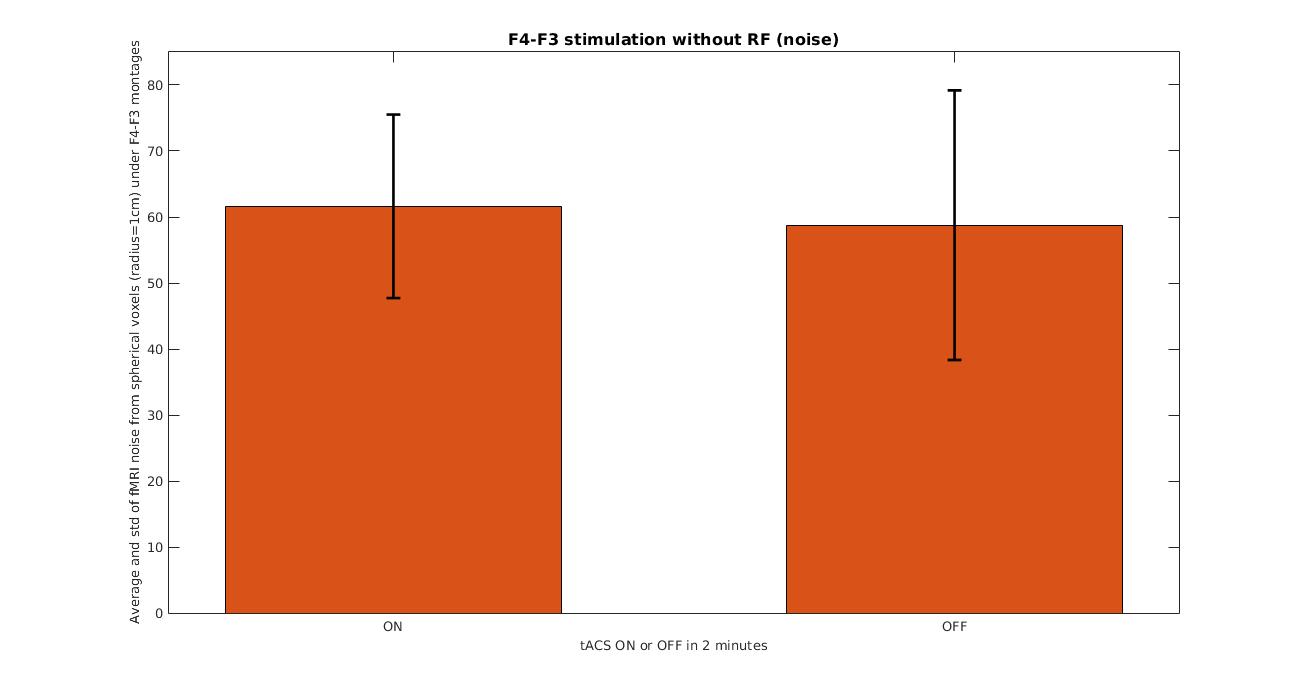
The average and standard deviation of fMRI signals from spherical voxels (radius=1cm) under
F4-F3 montages during tACS ON and OFF
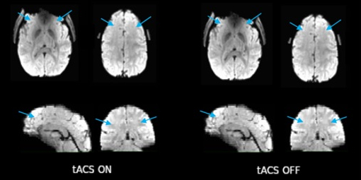
EPI images during tACS ON and OFF
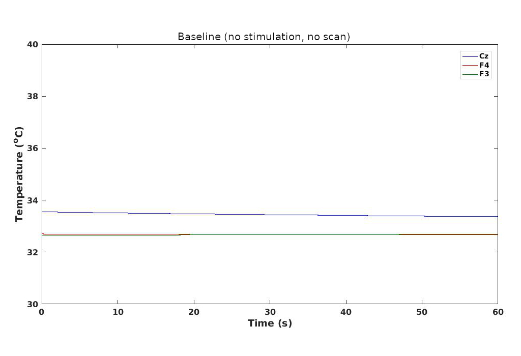
Baseline temperature on the scalp at F4, F3 and Cz electrodes when there is no stimulation and no scan
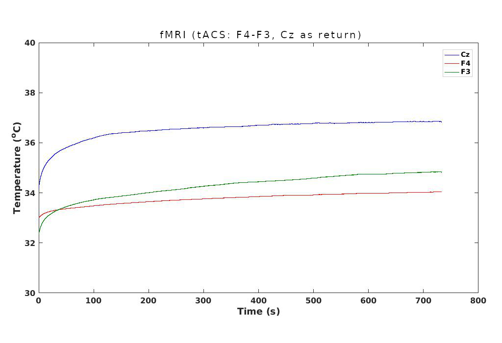
tACS stimulation: 2 mA, F4-F3, 2 min/2 min ON/OFF, 12 min