4146
Geometric accuracy of real-time tumor tracking and dynamic beam adaptation using the Australian MRI-Linear accelerator1Image X Institute, The University of Sydney, The University of Sydney, Australia, 2Department of Medical Physics, Ingham Institute for Applied Medical Research, Liverpool, Australia
Synopsis
A real-time tumor tracking system has been developed for MRI-guided radiation therapy. This system allows for tumor motion to be monitored during radiotherapy and for the radiation beam to be modified to compensate for this motion. The tumor location was found during treatment through the use of a template matching algorithm and 2D cine-MR images. The tumor location information was sent to a tracking algorithm that modifies the shape of the beam in real-time to ensure the tumor target is irradiated. The tumor tracking system has been tested and found to be of submilimeter accuracy.
Introduction
MRI-Linear accelerators (MRI-Linacs) are a new technology that is capable of MRI imaging prior to and during the delivery of radiation therapy1. Compared to conventional linear accelerators equipped with X-ray imagers, MRI-Linacs provide imaging with improved tissue contrast and functional information without additional radiation dose. MRI-Linacs enable treatment techniques that are difficult or not possible with X-ray imaging. One example is real-time MLC tracking where the radiation beam is modified during treatment delivery in response to tumor motion2. Tumor tracking ensures that radiation is delivered to the tumor and nearby healthy tissue is spared. This study describes the development and geometric characterization of a real-time tumor tracking system for the Australian MRI-Linac.Methods
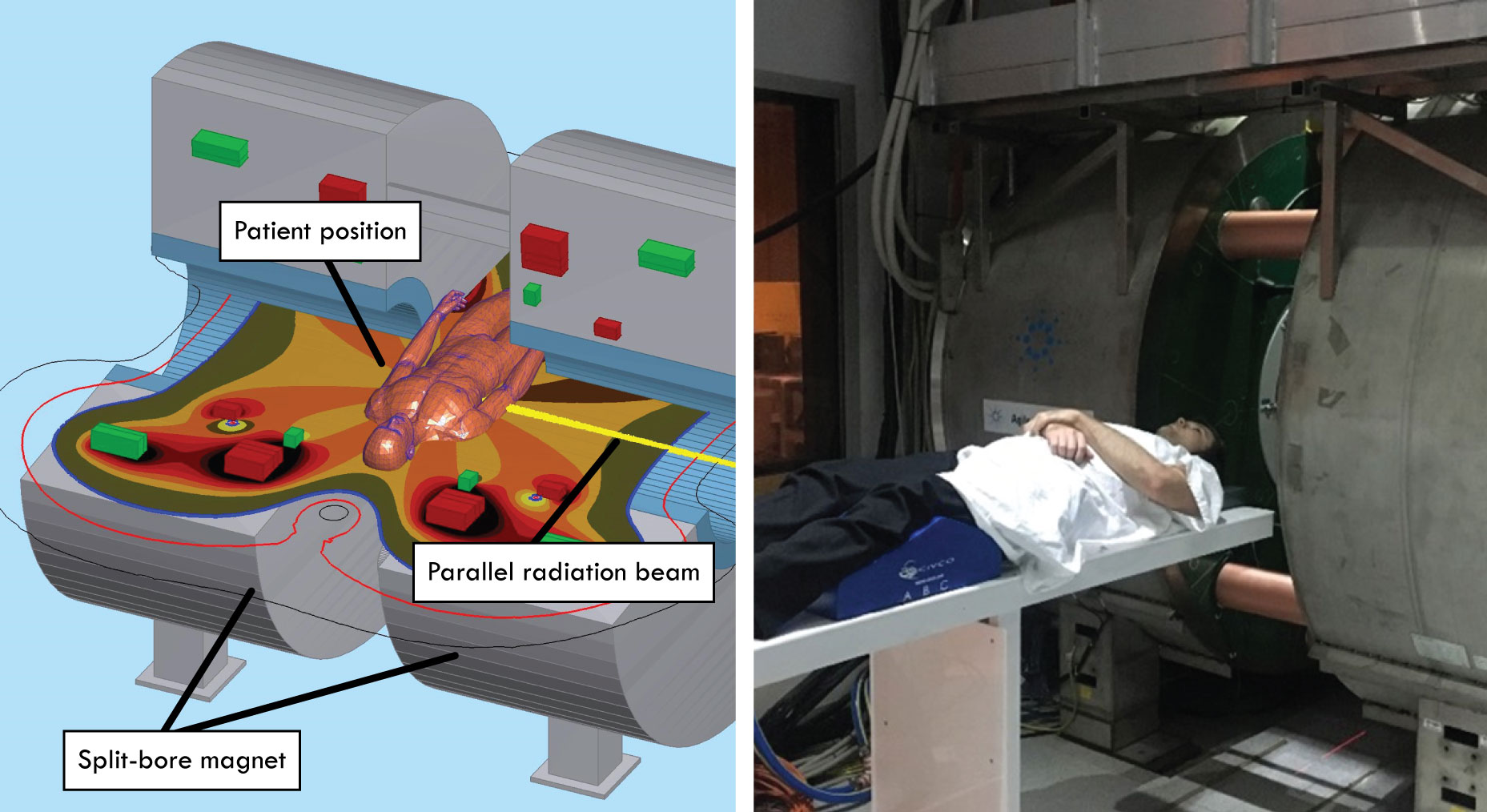
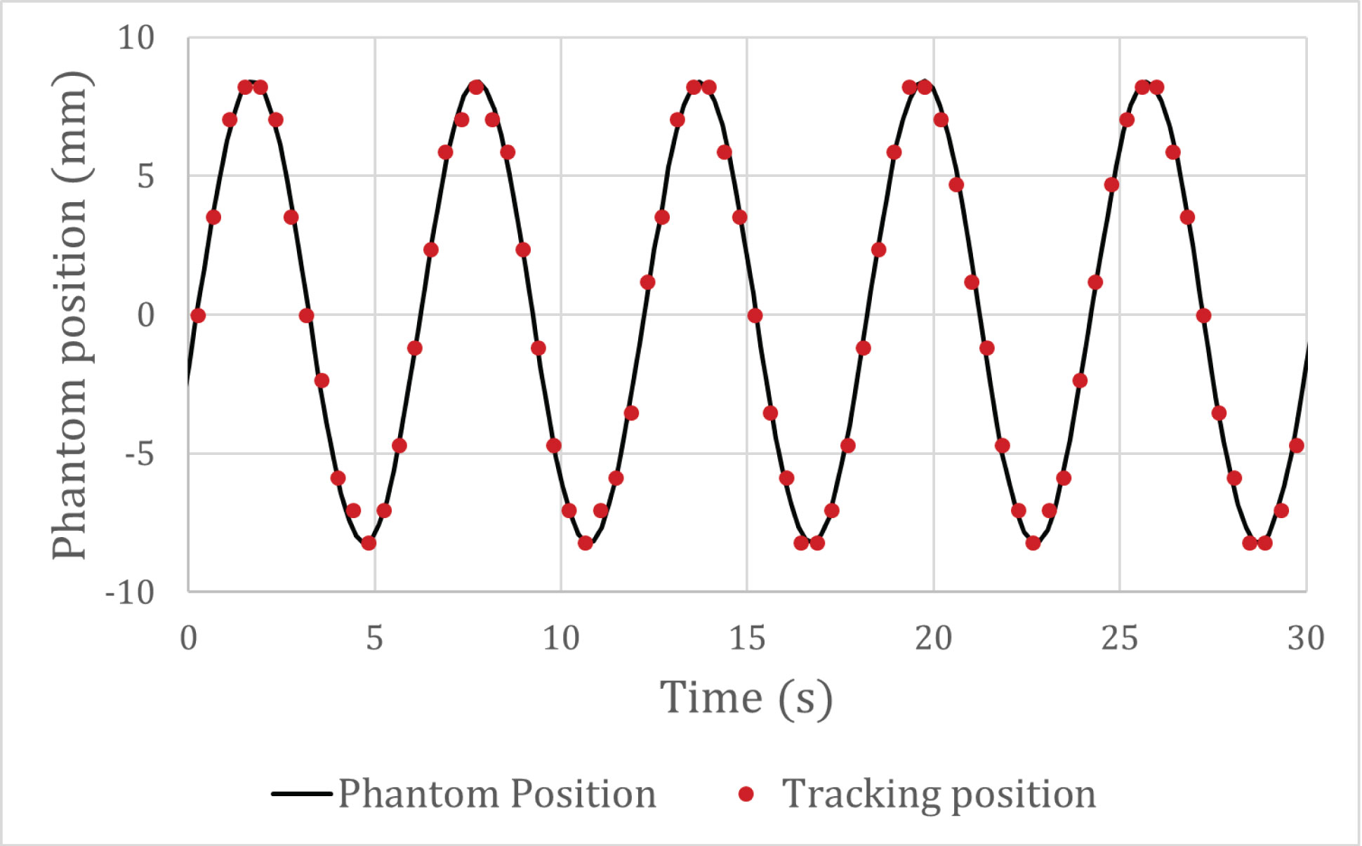
The Australian MRI-Linac, shown in Figure 1, is a prototype MRI-Linac that utilizes a 1 T split-bore magnet. The real-time target tracking system has two main components: (1) an imaging component that calculates the position of a selected target in 2D cine-MRI images acquired during radiation delivery and (2) a multi-leaf collimator (MLC) tracking component that dynamically reshapes the radiation beam aperture to compensate for motion.The imaging component is based on template matching algorithms published in the open-source OpenCV library. A region-of-interest (for example, the treatment target or a nearby surrogate structure) was selected as a template then matched in images acquired from the MRI-Linac during treatment. Images were acquired at a rate of 4 Hz and were upscaled using bicubic interpolation. The performance of the imaging component was experimentally verified using the Qasar Programmable Respiratory Motion Platform (Modus QA), programmed with sinusoidal motion. The tracked position of a 35 mm diameter spherical phantom was compared to the ground truth output of the motion phantom to analyze geometric accuracy.
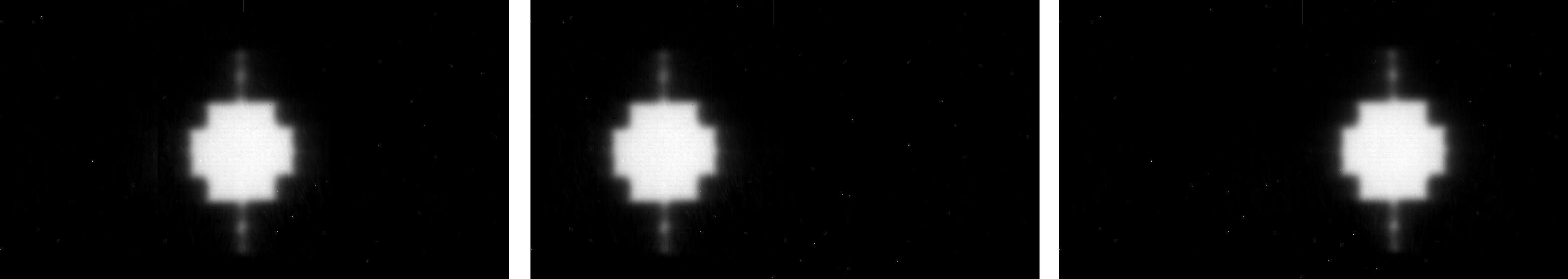
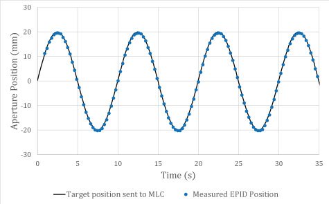
The position of the target found using the template matching algorithm was sent to the MRI-Linac’s MLC tracking component. The MLC tracking algorithm, described in Sawant et al3, recalculates the position of the 120 tungsten leaves within the MLC that define the shape of the radiation beam. The geometric accuracy of MLC tracking algorithm was measured using an EPID (electronic portal imaging device) that captures images of the radiation beam, shown in Figure 2.
Results
Figure 3 shows the geometric accuracy of the imaging component. The mean difference between the position of the motion phantom from the tracking algorithm compared to the position reported by the motion phantom was 0.04 ± 0.36 mm. The difference ranged from -0.76 mm to 0.82 mm. The tracking accuracy was limited by the spatial resolution of the images, originally 2.34 mm and interpolated to have a sub-pixel resolution of 1.17 mm.Figure 4 shows the geometric accuracy of the MLC tracking component. The mean difference position of the MLC leaves captured from the EPID compared to the position of target sent to the MLC was -0.32 ± 0.16 mm. The difference ranged from -0.71 mm to 0.21 mm.
Discussion
MLC tracking has been clinically implemented for lung and prostate cancer patients4,5 and has been shown to account for target motion, rotation and deformation6. MRI imaging is an ideal technology to improve MLC tracking as it can provide more detailed anatomical information. The real-time target tracking system developed in this work was found to accurately track the spherical target with submillimeter accuracy for both imaging and MLC tracking components.To test the performance of the system with clinical images, the template matching algorithm was also tested with images of rats taken on the Australian MRI-Linac and images of humans from a 3 T MRI. The tracking system was found to accurately track targets with appropriately selected templates, however it can fail if the selected region-of-interest does not contain enough contrast. In clinical use, the correlation coefficient calculated for each image can be used to monitor the accuracy of tracking.
Acknowledgements
This work has been funded by the Australian National Health and Medical Research Council Program Grant APP1132471.References
1. Liney, Gary P., et al. "MRI-linear accelerator radiotherapy systems." Clinical Oncology 30.11 (2018): 686-691
2. Glitzner, Markus, et al. "MLC-tracking performance on the Elekta unity MRI-linac." Physics in Medicine & Biology (2019)
3. Sawant, Amit, et al. "Management of three‐dimensional intrafraction motion through real‐time DMLC tracking." Medical physics 35.5 (2008): 2050-2061
4. Booth, Jeremy T., et al. "The first patient treatment of electromagnetic-guided real time adaptive radiotherapy using MLC tracking for lung SABR." Radiotherapy and Oncology 121.1 (2016): 19-25.
5. Keall, Paul J., et al. "The first clinical implementation of electromagnetic transponder‐guided MLC tracking." Medical physics 41.2 (2014).
6. Ge, Yuanyuan, et al. "Toward the development of intrafraction tumor deformation tracking using a dynamic multi‐leaf collimator." Medical physics 41.6Part1 (2014): 061703.
Figures