4128
Development of T1-based Skull Thermometry using a 3D Spiral Ultra-Short Echo Time Sequence1University of Virginia, Charlottesville, VA, United States, 2Biomedical Engineering, University of Virginia, Charlottesville, VA, United States, 3Department of Imaging Sciences & Innovation, Geisenger, Danville, PA, United States, 4Radiology and Medical Imaging, University of Virginia, Charlottesville, VA, United States
Synopsis
Skull temperature monitoring is important for MR-guided focused ultrasound (MRgFUS), as the skull is highly absorptive to acoustic energy. T1 thermometry uses T1 mapping to observe a linear increase in T1 with temperature but requires long acquisitions. Here we compare T1-weighted thermometry with T1 thermometry using a 3D spiral ultra-short-echo time sequence and show that T1 thermometry is feasible, but requires further acceleration, whereas T1-weighted thermometry results are nonlinear and inconsistent.
Introduction
In MRgFUS, acoustic waves are focused through the skull to destroy target brain tissue as a treatment for movement disorders. However, the skull can absorb up to 80% of the applied acoustic energy1. Despite current clinical precautions such as circulating cold water around the scalp and predicting the cooling time needed for the skull between sonications from a model, a recent study has shown that MRgFUS led to unintended skull lesions in 7 out of 30 patients2. Current precautions are also insufficient for skull heating from off-center targets, which limits MRgFUS application to brain therapies. Furthermore, the cooling time estimate is not patient specific and can thus prolong an expensive, uncomfortable treatment needlessly. Thus, there is a need for skull thermometry.Skull thermometry is challenging due to the extremely short T2* of cortical bone precluding standard proton resonance shift methods and because of the need for rapid imaging over a large FOV. Other researchers have shown a linear temperature dependence of T1 relaxation in cortical bone3. Our goal was to compare T1-weighted thermometry (1 image/1 temperature point) with T1-thermometry (2 images/temperature point) to determine a rapid but feasible method to image skull heating. Using a non-selective ultra-short-echo-time (UTE) 3D spiral sequence, we demonstrate that rapid T1-thermometry is feasible, and that it is more repeatable, and quantitative than T1-weighted thermometry.
Methods
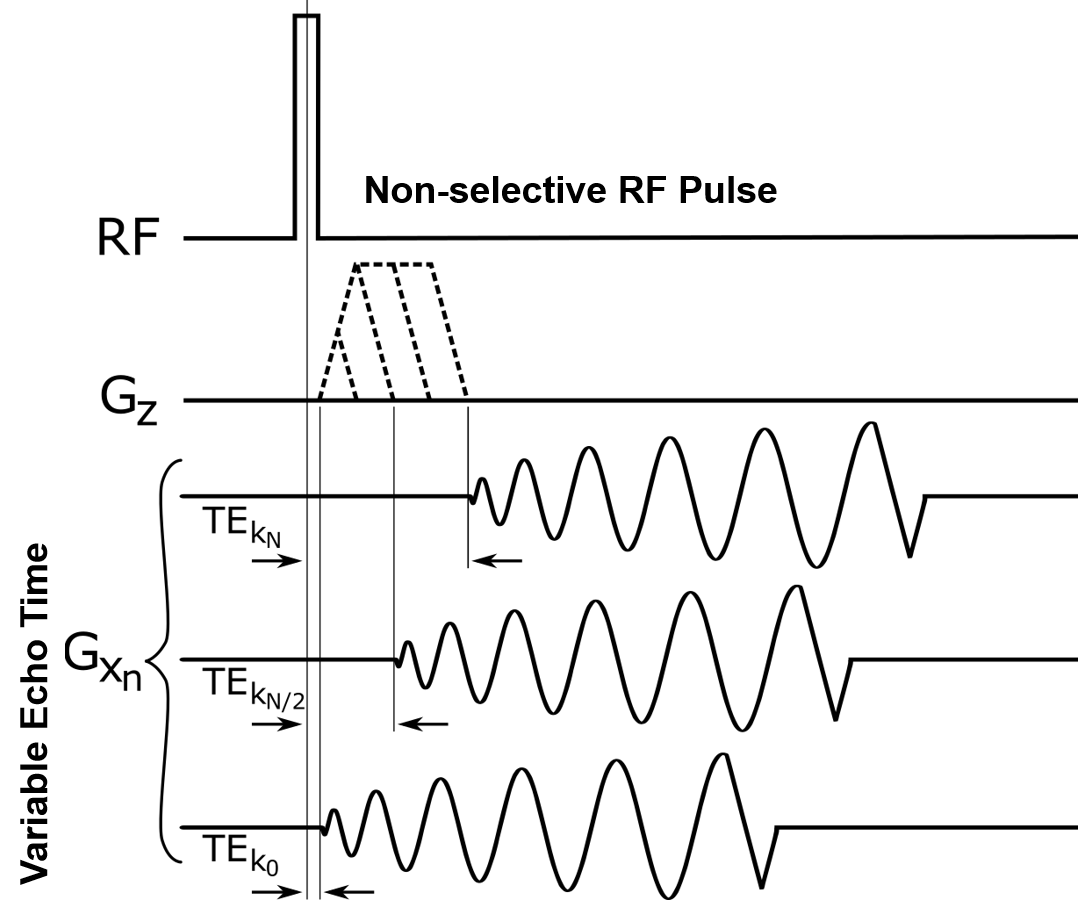
Two types of experiments were performed. For both, bovine femur bones were obtained from a butcher and stored in refrigeration before the experiment. Fiberoptic thermocouples were placed in a burr hole drilled into the cortical layer of each sample and in the water bath. In cooling experiments (2 trials), as shown in Fig. 1a, each bone was placed in water at ~60°C, equilibrated for 10 min., and then imaged with a 3T Siemens Prisma scanner with a single channel receiver coil hugging the sample at two optimal flip angles4 (dual angle T1-mapping) for every ~5°C of temperature loss. In heating experiments (3 trials) as shown in Fig. 1b, implemented for better temperature control, each bone was placed in a small lidded jar with undoped water. The small jar was placed into a bigger cylinder in which water from a water heater circulated. This ensured gradually changing temperature in the bone. During analysis, an ROI was selected in the bone. The mean signal in the ROI was measured at each flip angle. Temperature was then estimated using least squares regression of the standard spoiled GRE equation5.The 3D spiral-based UTE sequence as shown in Fig. 2 has a 3D stack-of-spirals readout. Each spiral readout occurs immediately after the through-plane phase-encoding gradient waveform is completed, resulting in a variable TE in the through-plane direction6. The sequence parameters were as follows: TE/TR 80us/11.3ms; FA: 8°, 35°; Voxel Size: 1x1x4mm3; Spiral Interleaves/Readout Duration: 203/0.5ms; Matrix Size: 160x160x22, TA per FA: 130s.
Results
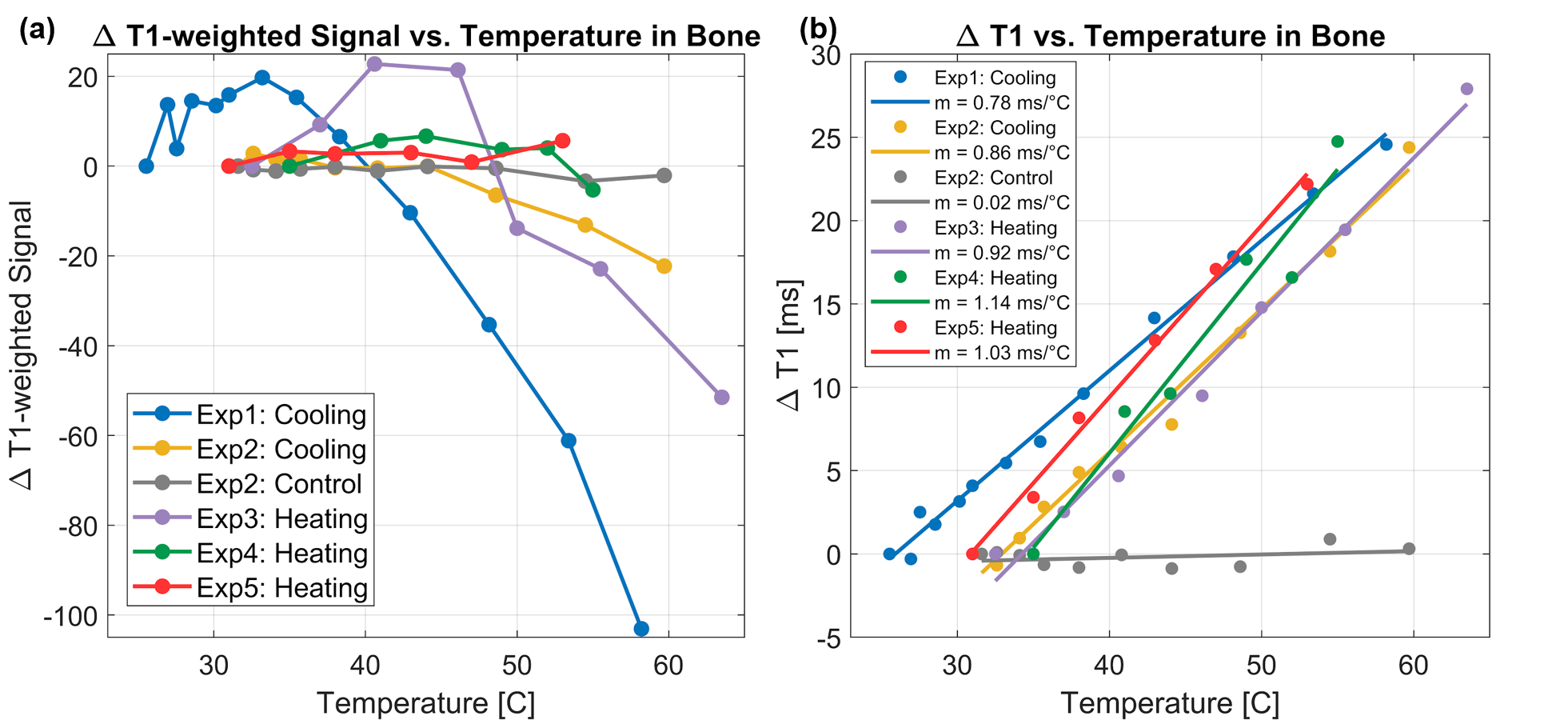
The T1-weighted signal at the 35° flip angle did not change linearly with temperature as shown in Fig. 3a, thus discouraging further pursuit of fast T1-weighted thermometry. However, the corresponding T1 measurements from the same ROI, but with an additional flip angle (FA: 8°) as shown in Fig. 3b, were strongly correlated with temperature. The average slope was 0.98 +/- 0.15 ms/°C, which is comparable to Han et al.’s result of 0.84 ms/°C measured using a slower 3D radial UTE pulse sequence3.Discussion and Conclusion
Skull thermometry must be fast, volumetric, and repeatable in its ability to show skull temperature’s return to baseline after sonication. Although the 35° flip angle T1-weighted method is fast, it is not linearly correlated with temperature, probably because of changes in T2* with temperature. In contrast, T1 mapping is strongly correlated and can be calibrated to indicate skull temperature. The 3D UTE, variable-density spiral sequence can be accelerated by decreasing resolution, in-plane undersampling, and through-plane partial Fourier reconstruction to meet the goal of 3D whole-skull thermometry in 90s or less. Development and validation are still needed to translate basic proof of concept into viable methods to monitor skull heating during transcranial MRgFUS.Acknowledgements
This research was partly supported by Siemens Medical Solutions and by the NIH Biomedical Data Science Training Grant (5T32LM012416-04). The authors acknowledge Josef Pfeuffer, Thomas Benkert, and Berthold Kiefer for their help with this project.References
1. Gianmarco Pinton, Jean-Francois Aubry, Emmanuel Bossy, Marie Muller, Mathieu Pernot, and Mickael Tanter. Attenuation, scattering, and absorption of ultrasound in the skull bone. Medical physics, 39(1):299–307, jan 2012. ISSN 0094-2405.
2. Schwartz, Michael L., et al. “Skull Bone Marrow Injury Caused by MR-Guided Focused Ultrasound for Cerebral Functional Procedures.” Journal of Neurosurgery, 2018, pp. 1–5., doi:10.3171/2017.11.jns17968.
3. Han, Misung, et al. “Assessing Temperature Changes in Cortical Bone Using Variable Flip-Angle Ultrashort Echo-Time MRI.” Magn Res Med. 2015 Dec; 74(6): 1548–1555.
4. Deoni, S. C., Rutt, B. K. and Peters, T. M. (2003), Rapid combined T1 and T2 mapping using gradient recalled acquisition in the steady state. Magn. Reson. Med., 49: 515-526. doi:10.1002/mrm.10407
5. Nishimura, Dwight George. Principles of Magnetic Resonance Imaging. Dwight G. Nishimura, 2016.
6. Fielden S. et al. ISMRM 2015;23: p3867.
7. Odéen, Henrik, and Dennis L. Parker. “Non-Invasive Thermometry with Magnetic Resonance Imaging.” Theory and Applications of Heat Transfer in Humans, 2018, pp. 267–299., doi:10.1002/9781119127420.ch15.
8. Odeen O., Bolster B., Jeong E. Parker D. “Investigation of temperature dependent changes in signal intensity, T1 and T2* in cortical bone”. Abstract. In:ISMRM 2016.
9. Miller GW. “MR bone Imaging.” J Ther Ultrasound. 2015; 3(Suppl 1): O37.
10. Odéen, Henrik, and Dennis L. Parker. “Non-Invasive Thermometry with Magnetic Resonance Imaging.” Theory and Applications of Heat Transfer in Humans,2018, pp. 267–299., doi:10.1002/9781119127420.ch15.
11. Ramsay, Elizabeth, et al. “Temperature-Dependent MR Signals in Cortical Bone: Potential for Monitoring Temperature Changes during High-Intensity Focused Ultrasound Treatment in Bone.” Magnetic Res Med, vol. 74, no. 4, 2014, pp. 1095–1102., doi:10.1002/mrm.25492.
Figures