4121
Focused Ultrasound Enhances Connectivity of Default Mode Network in Rat Brain1National Tsing Hua University, Hsinchu, Taiwan, 2Chang Gung University, Taoyuan, Taiwan
Synopsis
Focused ultrasound is a noninvasive, deep-penetrating, and spatially well-defined modality to modulate the neural activity. In this study, we used resting state functional MRI (rs-fMRI) to investigate the functionally connectivity after stimulation in the brain. Rats underwent focused ultrasound stimulation targeted unilaterally to the ventral posteromedial and ventral posterolateral thalamic nuclei (VPM/VPL) in the left thalamic region. The default mode network (DMN) showed hyper-correlation after sonication, and as time proceed, the correlation has a tendency to decline slowly in about 3 days.
Introduction
Neuromodulation is an emerging technology that induce therapeutic alteration of the nervous system by delivering energy to a targeted area. [1] Focused ultrasound (FUS) is a noninvasive, deep-penetrating, and spatially well-defined modality to deliver energy to central nervous system. Instead of monitoring the transient neural activation at the localized sonication region, we proposed to observe the neuromodulation effect on the change of connectivity of default mode network (DMN). Therefore, in this study, resting state functional MRI (rs-fMRI) was performed as a noninvasive tool to monitor the DMN of rat brains.Methods
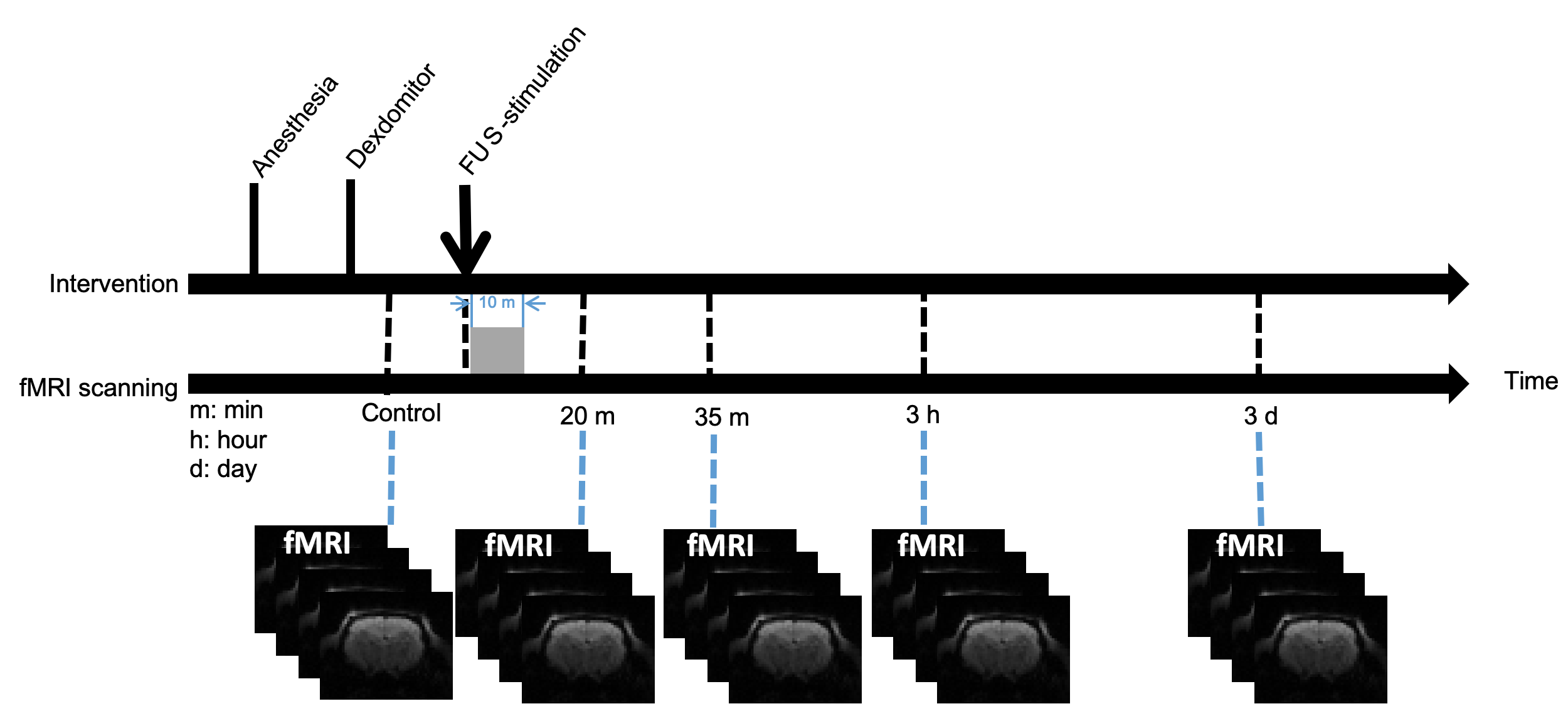
All protocols were approved by local IACUC. Adult male Sprague-Dawley rats (350–420 g) were used in this study. MRI experiments were performed on 7-Tesla Bruker ClinScan scanner. Anatomical images were obtained using turbo-spin-echo (TSE) with scanning parameters of TE/TR = 14ms/4000ms, FOV = 30 × 30 mm2, matrix size = 256 × 256, slice thickness = 1 mm, number of averages = 2. For functional scans, 300 consecutive volumes with 15 coronal slices were acquired using gradient echo EPI with TE/TR = 20 ms/1000 ms, FOV = 30 × 30 mm2, matrix size = 64 × 64, and slice thickness = 1 mm. Animals were anesthetized with 2% isoflurane mixed with O2 at flow rate of 1L/minute for inserted a catheter into the tail vein and fixed in place for intravenous injection. The isoflurane was immediately disconnected after intravenous injection 0.05 mg/kg Dexdomitor® and left to recover from isoflurane for 15 minutes. FUS was targeted to the ventral posteromedial and ventral posterolateral thalamic nuclei (VPM/VPL) region in left thalamus. Burst-mode focused ultrasound was delivered with pulse repetition frequency =100 Hz, spatial peak negative pressure amplitude = 0.25 MI, duty cycle = 30%, and the stimulation time was 30 seconds on and 90 seconds off for 5 cycles. 5 data sets (N=7) of rs-fMRI were collected at different time-point (figure 1): control group without FUS stimulation, 20 minutes after FUS, 35 minutes after FUS, 3 hours after FUS, and 3 days after FUS.Results
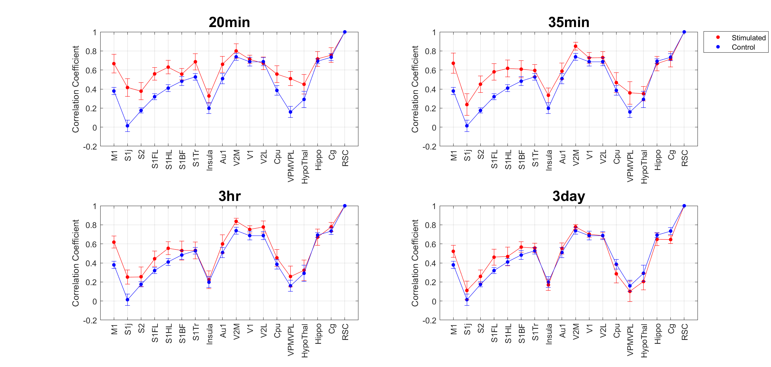
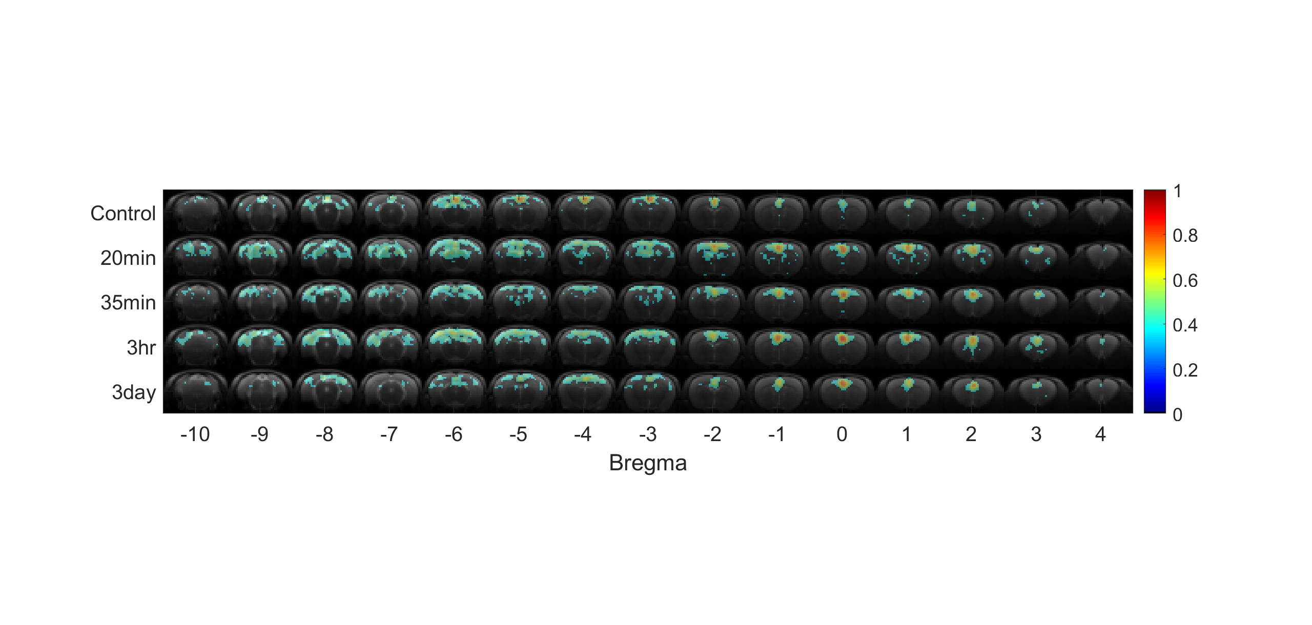
Line charts of connectivities between RSC and different brain regions were shown in figure 2. Compared to the control group, it is noted that the line of correlation coefficients was elevated in regions including VPM/VPL, M1, S1, and S2 in both data sets of 20 and 35 minutes after FUS. Figure 3 showed that the spatial distribution of seed- mapping DMN was enlarged after FUS, which may imply the enhancement of synchronization after FUS. The observed enhancement of DMN was decayed with time, and the averaged connectivities were almost identical between the control group and 3 days after FUS.Discussions
Although VPM/VPL were not include in the DMN, the result suggests the focal stimulation in a thalamic region can lead to non-local alterations of resting state functional connectivity, which may be contributed by the interaction between neural networks. Our FUS stimulations raised the synchronization in brain regions, and therefore enlarged the spatial distribution of DMN. As time proceeds, the inter-brain region's connectivity gradually reverted close to the baseline in 3 days, which implied the impact of sonication affect was temporal.Conclusion
Our study successfully demonstrated that combining rs-fMRI with FUS could be a powerful platform for future neuromodulation studies. Reversible hyper-connectivity of DMN can be induced by FUS and then monitored by rs-fMRI longitudinally. We expected that the enhancement of neural connection strength potentially benefits on psychiatric disorder and neurodegenerative diseases.[2] Patients with Parkinson's disease [3], Alzheimer's disease, and depression have lower connectivity of DMN, and enhancing the connectivity may be an alternative treatment method.Acknowledgements
We thank the instrument support from Center for Advanced Molecular Imaging and Translation, Chang Gung Memorial Hospital, Linkou.References
1. Todd, N., et al., Focused ultrasound induced opening of the blood-brain barrier disrupts inter-hemispheric resting state functional connectivity in the rat brain. Neuroimage, 2018. 178: p. 414-422.
2. Min, B.K., et al., Focused ultrasound-mediated suppression of chemically-induced acute epileptic EEG activity. BMC Neurosci, 2011. 12: p. 23.
3. Baudrexel, S., et al., Resting state fMRI reveals increased subthalamic nucleus-motor cortex connectivity in Parkinson's disease. Neuroimage, 2011. 55(4): p. 1728-38.
Figures