4104
A Wearable and Flexible 23Na Transmit/Receive Breast Coil at 3T1The Graduate School of Natural and Applied Sciences, Dokuz Eylul University, Izmir, Turkey, 2Institute of Biomedical Engineering, Bogazici University, Istanbul, Turkey, 3Department of Radiology and Medical Informatics, University of Geneva, Geneva, Switzerland, 4Department of Electrical and Electronic Engineering, Ege University, Izmir, Turkey
Synopsis
A novel wearable and flexible 23Na transmit/receive breast coil made from a highly conductive thread was introduced for 3 T. Comparison studies were conducted with a 23Na traditional breast coil having the same geometry as the flexible one. Sensitivity maps of 23Na traditional breast coil and 23Na flexible textile breast coil were similar. In addition, 23Na flexible textile breast coil provided better SNR performance when different NaCl concentration were to be imaged. In conclusion, applicability of textile coils on 23Na MRI was demonstrated by means of similar SNR results of 23Na flexible textile breast coil and 23Na traditional breast coil.
Introduction
Biomedical applications of sodium (23Na) MRI in human body involves brain, breast, heart, skeletal muscle, cartilage, abdominal, and cancer imaging1. 23Na MRI of breast is particularly used in diagnosis of breast cancer and determining its stage, as well as monitoring treatment response2,3. Fixed-sized breast coils used in clinical applications could lead to difficulties in quality factor, hence, might indirectly result in degradation of image quality or discomfort for patients because of the squeezing sensation. A previous study4 validated the convenience and usability of a conductive textile-based proton breast coil. In this study, a novel single channel wearable and flexible 23Na transmit/receive breast coil is introduced for 3T.Methods
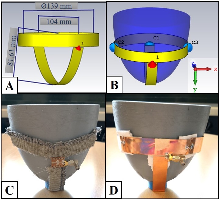
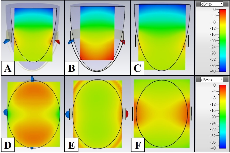
Simulation StudiesSimulation studies were conducted in CST (Computer Simulation Technology, AG, USA). The geometry of the breast coil (Fig 1.A,B) was determined so that it would create a magnetic field in both the sagittal plane and the coronal plane. X, Y, and Z directions in the simulation indicated the sagittal plane, the coronal plane, and B0, respectively. Magnetic field distributions (B1 field) of selected slices are given in Fig 2.
23Na Flexible Textile Breast Coil
A highly conductive silver thread (Statex, DE, USA) was determined as the material of the textile coil. 14 cm of textile and 12 cm of textile were cut to create the top ring, whereas, 10 cm of textile was used to create the bottom ring (Fig 1.C). Since the circuit components could not be soldered directly on the textile, a connection circuit was designed for this purpose. Three serially connected capacitors (220pF, 270pF, ATC Corp., USA) were used to tune the textile coil. The matching circuit was designed by a serially connected inductor (39nH, CoilCraft, 0603HP, USA) and a capacitor (390pF, Johanson Technology, EIA 1111, USA) connected in parallel, such that |S11| was -12 dB at 32.6 MHz (Larmor frequency of sodium at 3 T).
23Na Traditional Breast Coil
A copper strip with a thickness of 0.3 mm and width of 20 mm was used to create a traditional breast coil (Fig 1.D). Three capacitors (220pF, 270pF, ATC Corp., USA) were connected serially to tune the coil. The matching circuit was achieved by a serially connected inductor (22nH, CoilCraft, 0603HP, USA) and two capacitors (470pF and 33pF, Johanson Technology, EIA 1111, USA) connected in parallel, thus |S11| was -12 dB at 32.6 MHz.
Phantom Studies
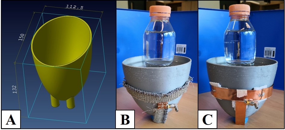
The shell of the phantom was printed by a 3D printer (Ultimaker2, NL) and was filled with 300 mM of NaCl solution to obtain sensitivity maps (reception profile) of the breast coils. Afterwards, the phantom was filled with 100 mM of NaCl solution along with a separate bottle of 300 mM of NaCl solution (Fig 3) to create contrast in MR images.
Tim and Flex Coil Interface of Siemens (Siemens Magnetom Prisma 3 T, Siemens Healthcare AG, DE) was used for 23Na imaging. Both 23Na flexible textile breast coil and 23Na traditional breast coil were used in transmit/receive mode. Flash scan parameters were: TR/TE = 100/3.3 ms, flip angle= 90°, voxel size= 4x4x20 mm, FOV= 320x320 mm, averages= 32 with a total scan time of 4 min. 80 V was applied to obtain 90° flip angles for both breast coils.
Image Analysis
Acquired images were analysed with BreastIS5 post-processing and analysis software to assess and compare their quality. Coil inhomogeneity effects were corrected using coil sensitivity maps and image SNR was calculated for both original and coil inhomogeneity corrected images.
Results
Phantom ResultsThe maximum values of the resultant FID peak of 23Na traditional breast coil and 23Na flexible textile breast coil were 576 and 507, respectively.
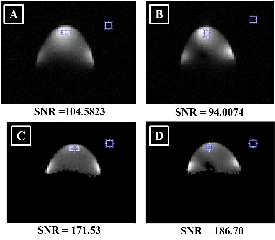
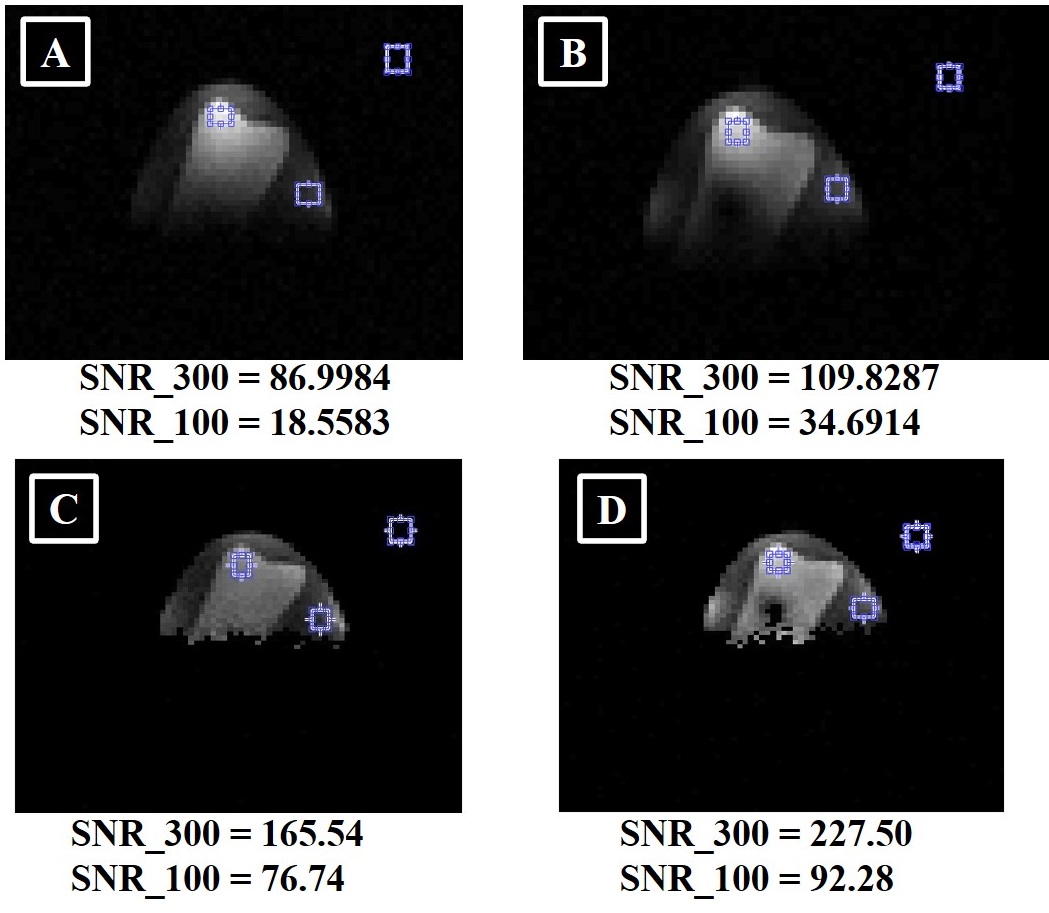
Sensitivity maps (Fig 4) of 23Na traditional breast coil and 23Na flexible textile breast coil were similar. While uncorrected SNR of 23Na traditional breast coil was higher, 23Na flexible textile breast coil had better corrected SNR. 23Na flexible textile breast coil provided better contrast in two different solution setup (Fig 5). Fig 4.A and Fig 4.B were similar to Fig 4.C and Fig 4.D in terms of SNR results, including only signal areas in Fig 4.B and Fig 4.D. SNR_300 was almost three times higher than SNR_100 in Fig 5.D as expected, yet this could not be observed evidently in Fig 5.C. I would not specify that. It is obvious.
Discussion
Measured sensitivity maps of both 23Na flexible textile breast coil and 23Na traditional breast coil were similar to simulated B1 fields. Signal loss in areas away from the coil and sensitivity in areas where capacitors were placed or the feeding point existed were expected. However, 23Na flexible textile breast coil provided better SNR performance (Fig 5.B and Fig 5.D) when different NaCl concentration were to be imaged.In conclusion, applicability of textile coils on 23Na MRI was demonstrated by means of similar SNR results of 23Na flexible textile breast coil and 23Na traditional breast coil.
Acknowledgements
We thank Center for Biomedical Imaging (CIBM, Switzerland) for possibilities provided for working at RF Lab and 23Na MRI acquisition.
This study was partly funded by TUBITAK (The Scientific and Technological Research Council of Turkey) under grant no. 116E155 and 2214-A International Doctoral Research Fellowship Programme.
References
1. Madelin, G., et al., Sodium MRI: Methods and applications. Progress in Nuclear Magnetic Resonance Spectroscopy, 2014. 79: p. 14-47.
2. Ouwerkerk, R., et al., Elevated tissue sodium concentration in malignant breast lesions detected with non-invasive Na-23 MRI. Breast Cancer Research and Treatment, 2007. 106(2): p. 151-160.
3. Jacobs, M.A., et al., Multiparametric and multinuclear magnetic resonance imaging of human breast cancer: Current applications. Technology in Cancer Research & Treatment, 2004. 3(6): p. 543-550.
4. Kahraman Agir, B., Bayrambas B., Yegin K., Ozturk Isik E., Wearable and Stretchable Surface Breast Coil, in 28th Scientific Meeting, International Society for Magnetic Resonance in Medicine. 2019. Montreal, 3782.
5. Bayrambas B., Hatay G.H., Yegin K., Ozturk-Isik E., BreastIS: An Analysis Software for Magnetic Resonance Image Analysis of Breast, in 36th Scientific Meeting, European Society for Magnetic Resonance in Medicine and Biology. 2019. Rotterdam, 1469.
Figures