4102
Highly-Flexible Highly-Decoupled 1H RF Coils for MR imaging of the lungs as utilized within a 129Xe Transmit-Receive RF Coil at 1.5 T1University Of Sheffield, Sheffield, United Kingdom, 2GE Healthcare, Aurora, OH, United States
Synopsis
Flexibility of a RF coil constructed with flexible printed circuit board is reduced when it is co-housed with another RF coil, as typically applied for multinuclear MR imaging. In this work, for multinuclear 129Xe-1H lung imaging application, we propose to use highly-flexible multiple linked resonators for 1H RF coil along with 129Xe RF coil. These highly-flexible 1H RF coils are also highly-decoupled at 129Xe Larmor frequency. For the demonstration of multi-nuclear lung imaging, sixteen receive only highly-flexible highly-decoupled 1H RF coils were fixed on to a transmit-receive 129Xe RF coil, and 129Xe and 1H lung images were obtained.
Introduction
Obtaining high quality 1H lung MR images during multinuclear lung MR imaging with hyperpolarized (HP) gases such as 3He and 129Xe is of research and clinical interest1-6, because it would enable accurate quantification of lung structure and function in the same scan session4-6. High quality 1H lung MRI is hindered due to low proton density and short transverse relaxation times5-7. One approach to improve the image quality of 1H lung MRI in the same scan session as a HP gas MR exam would be to integrate a 1H RF coil array with the non-proton (129Xe, 3He) RF coil, wherein they are electrically isolated5,6. However, nesting two or more RF coils, where all the RF coils are constructed using printed circuit boards or equivalent as described in previous literature6, increases bulk of the coil, reduces the flexibility and thereby is less conforming to the human body, which can reduce the overall image quality. Although polyamide based printed circuit boards can be made very flexible, capacitors and their supporting structure brings rigidity to the array design. Taking advantage of recent advances of highly-flexible highly-decoupled (HFHD) RF coils, commercially called as AirTM is our proposed solution to this issue8. Such coils are designed based upon the construction of multiple linked resonator elements. Previous investigations have shown that the ratio of unloaded to loaded Q factor, which determines the overall sensitivity of the RF coil, was comparable with typical/conventional RF coils9. In this work, we investigate the feasibility of using HFHD 1H loops nested within a xenon transmit-receive RF coil for multi-nuclear lung imaging.Method
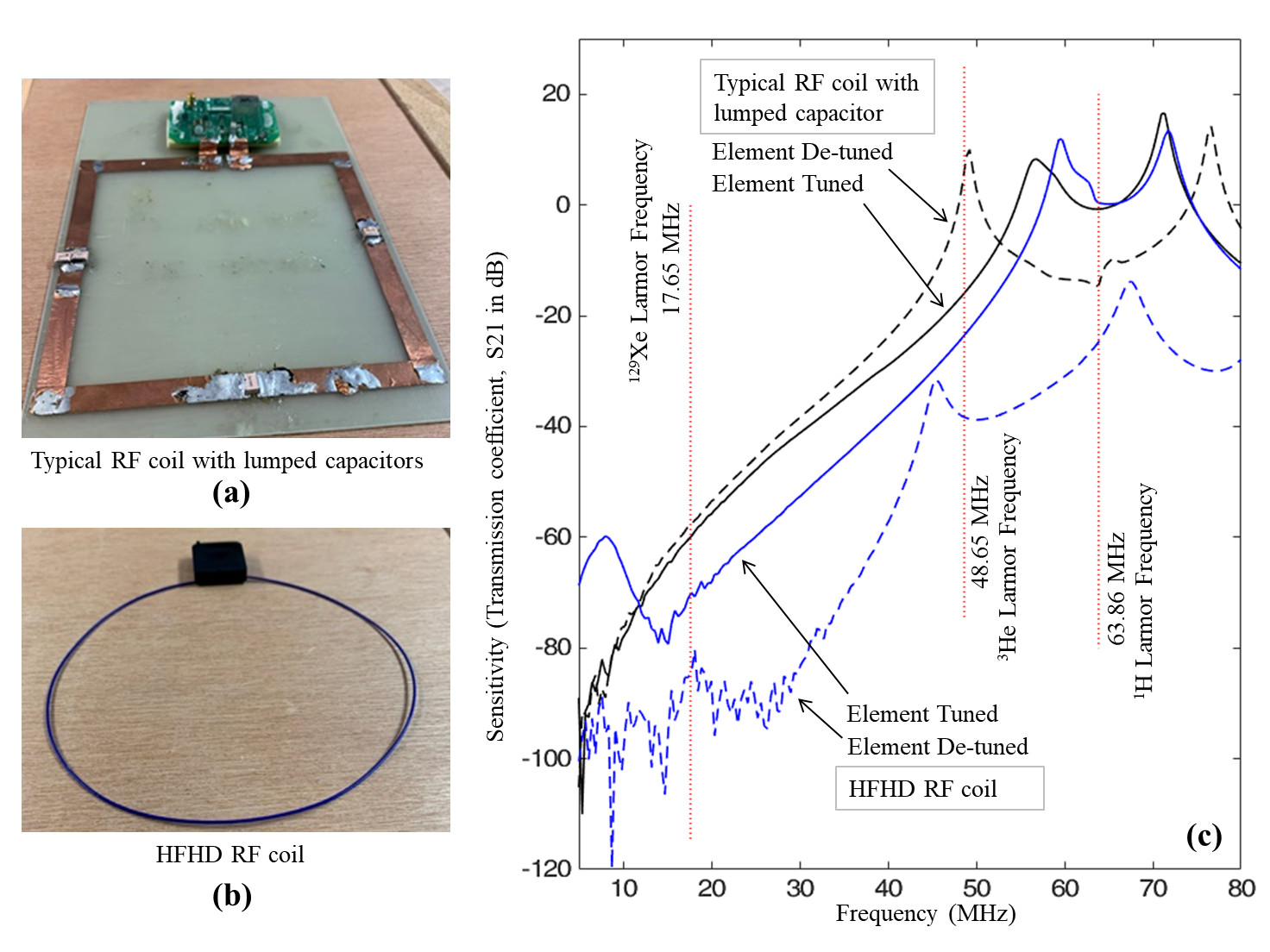
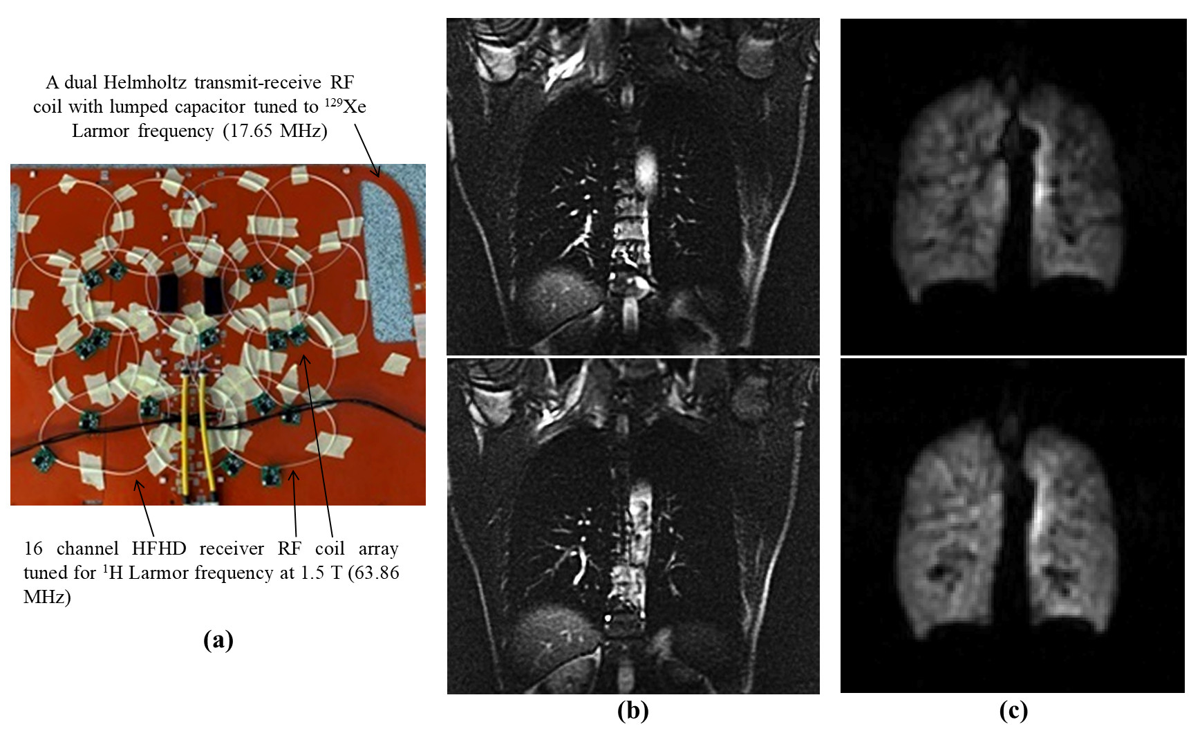
The feasibility of co-housing HFHD RF Coils tuned to 1H Larmor frequency inside a RF coil tuned to a non-proton Larmor frequency was assessed by comparing the intrinsic electrical isolation with a typical RF coil element with lumped capacitors. In a RF laboratory, this was assessed by comparing the relative sensitivity or power level at non-proton to 1H Larmor frequencies. Sixteen HFHD elements were built with a diameter of 11 cm from components (GE Coils Aurora ,OH, USA) and optimised for 63.86 MHz. A typical/conventional RF coil loop with lumped capacitors was constructed as shown in Figure 1(a) and was compared with flexible coil of similar spatial dimension as shown in Figure 1(b). The sensitivity of the RF coil was measured with the RF coil tuned and detuned, measured as transmission coefficient (S21 in dB). A dual Helmholtz transmit-receive RF coil was built on a polyamide substrate to image the inhaled HP 129Xe gas in the lung airspace. Sixteen flexible HFHD RF coils were fixed on to the 129Xe RF coil as shown in the Figure 2(a), such that the neighbouring elements were isolated using geometric overlap and preamplifier decoupling was used to isolate with all the other elements. 129Xe was hyperpolarized using spin exchange optical pumping polarizer (10), and 500 ml of HP 129Xe was mixed with 500 ml of N2 before administering the dose for ventilation scan. The imaging parameters were; balanced steady-state free precession, TE 2.2 ms, TR 6.7 ms, bandwidth 8 kHz, matrix 100 x 100, field-of-view 48 cm, slice thickness 10 mm. HP 129Xe imaging was performed on a GE Signa HDx 1.5 T scanner. Imaging parameters for 1H lung MR imaging were; balanced steady-state free precession, TE 0.8 ms, TR 2.7 ms, bandwidth 125 kHz, matrix 256 x 192, field of view 48 cm, slice thickness 10 mm. 1H imaging was performed on GE MR450w.Results
As seen in Figure 1(c), for a typical RF coil, when tuned, the relative sensitivity is moderate and low at 3He and 129Xe frequencies respectively. However, when detuned, the sensitivity at 3He frequency drastically increases and at 129Xe frequency remains unchanged. In contrast, the HFHD coil has significantly lower sensitivity when detuned. In addition, asymmetric frequency response can be observed by comparing the sensitivity of HFHD coils between tuned and detuned state, as seen in Figure 1(c).Figure 2 (b) shows 1H images of lungs acquired with 16 channel flexible HFHD coils, and Figure 2 (c) shows HP 129Xe lung images acquired with flexible transmit-receive RF coil.
Discussion
Conventionally, in order to mitigate the coupling between 1H and non-proton RF coils, trap circuits are often used6,11, which are bulky and generate heat. HFHD 1H RF coil elements at 1.5 T shows significant decoupling performance at 129Xe and 3He Larmor frequencies without using traps. As a receiver, HFHD coil elements not only exhibits similar sensitivity at 1H Larmor frequency as compared to typical/conventional RF coils but also has much reduced sensitivity at 129Xe Larmor frequency. During transmit, when the HFHD coil is detuned, the decoupling is better throughout non-proton Larmor frequencies, contributed by the asymmetric frequency response observed between tuned and detuned state of the HFHD RF coil. Thus, by using HFHD coil elements, the need for additional traps or blocking circuits can be avoided. In contrast to previous attempts of nested RF coils in-situ for multi-nuclear lung imaging6, this proposed solution is more practical.Conclusion
This work demonstrates an arrangement of RF coils for multinuclear lung imaging by combining 129Xe transmit-receive coil and 1H HFHD RF coils.Acknowledgements
This work was funded by the Engineering and Physical Sciences Research Council (EPSRC - EP/D070252/1) and Medical Research Council (MRC - MR/M008894/1). Authors would like to thank Dashen Chu, Yun-Jeong Stickle and Guido Kudielka.References
1. Woodhouse N, Wild JM, Paley MNJ, Fichele S, Said Z, Swift AJ, Van Beek EJR. Combined helium-3/proton magnetic resonance imaging measurement of ventilated lung volumes in smokers compared to never-smokers. Journal of Magnetic Resonance Imaging 2005;21(4):365-369.
2. Zheng J, Leawoods JC, Nolte M, Yablonskiy DA, Woodard PK, Laub G, Gropler RJ, Conradi MS. Combined MR proton lung perfusion/angiography and helium ventilation: Potential for detecting pulmonary emboli and ventilation defects. Magnetic Resonance in Medicine 2002;47(3):433-438.
3. Wild JM, Ajraoui S, Deppe MH, Parnell SR, Marshall H, Parra-Robles J, Ireland RH. Synchronous acquisition of hyperpolarised 3He and 1H MR images of the lungs - maximising mutual anatomical and functional information. NMR in Biomedicine 2011;24(2):130-134.
4. Wild JM, Marshall H, Xu X, Norquay G, Parnell SR, Clemence M, Griffiths PD, Parra-Robles J. Simultaneous imaging of lung structure and function with triple-nuclear hybrid MR imaging. Radiology 2013;267(1):251-255.
5. Rao M, Wild JM. RF instrumentation for same-breath triple nuclear lung MR imaging of (1)H and hyperpolarized (3)He and (129)Xe at 1.5T. Magn Reson Med 2016;75(4):1841-1848.
6. Rao M, Robb F, Wild JM. Dedicated receiver array coil for 1H lung imaging with same-breath acquisition of hyperpolarized 3He and 129Xe gas. Magnetic Resonance in Medicine 2015;74(1):291-299.
7. West JB. Respiratory physiology: the essentials: Lippincott Williams & Wilkins; 2012.
8. Vasanawala SS, Stormont R, Lindsay S, Grafendorfer T, Cheng JY, Pauly JM, Michael L, Scott G, Guzman JX, Taracila V, Chirayath D, Robb F. Development and Clinical implementation of very light weight and highly flexible Air technology arrays. Proc Intl Soc Mag Reson Med 25 P 0755 2017.
9. McGee KP, Stormont RS, Lindsay SA, Taracila V, Savitskij D, Robb F, Witte RJ, Kaufmann TJ, Huston J, 3rd, Riederer SJ, Borisch EA, Rossman PJ. Characterization and evaluation of a flexible MRI receive coil array for radiation therapy MR treatment planning using highly decoupled RF circuits. Phys Med Biol 2018;63(8):08nt02.
10. Norquay G, Collier GJ, Rao M, Stewart NJ, Wild JM. 129Xe-Rb Spin-Exchange Optical Pumping with High Photon Efficiency. Physical Review Letters 2018;121(15):153201.
11. Magill AW, Gruetter R. Nested Surface Coils for Multinuclear NMR. eMagRes: John Wiley & Sons, Ltd; 2007.
Figures