4101
Multinuclear RF coil for 13C and 1H breast MR imaging at 3.0 T1University Of Sheffield, Sheffield, United Kingdom, 2Memorial Sloan Kettering Cancer Center, New York, NY, United States
Synopsis
Hyperpolarized 13C MRI enables the investigation of metabolism of breast lesions for diagnosis and to monitor disease progression. Acquiring 13C MR images along with 1H MR images using a multi-nuclear RF coil enables accurate co-registration of the images, thereby providing complementary structural and functional images. In this work, we propose a design for multinuclear 13C-1H RF coil array for breast MR imaging, and assess the performance using both RF and MRI measurements. Both 13C and 1H MR images were obtained of using phantoms.
Introduction
Hyperpolarized (HP) 13C MRI enables the study of metabolic pathways which is vital to furthering our understanding of several diseases and their progression, such as cancer and other metabolic diseases1. Breast cancer is especially aggressive2,3, being able to investigate the metabolism of breast lesions using HP 13C MRI has potential to diagnose and monitor disease progression and treatment response4,5. To obtain high diagnostic quality MR images of HP 13C in vivo in the breast, several engineering aspects such as efficient polarization, sensitive RF coils and optimal pulse sequences are critical. In this work, we present a sensitive multinuclear RF coil for 13C and 1H imaging at 3.0 T, and providing a multinuclear RF coil which can enable registration of 13C images with 1H images for future functional and structural human breast MRI studies.Method
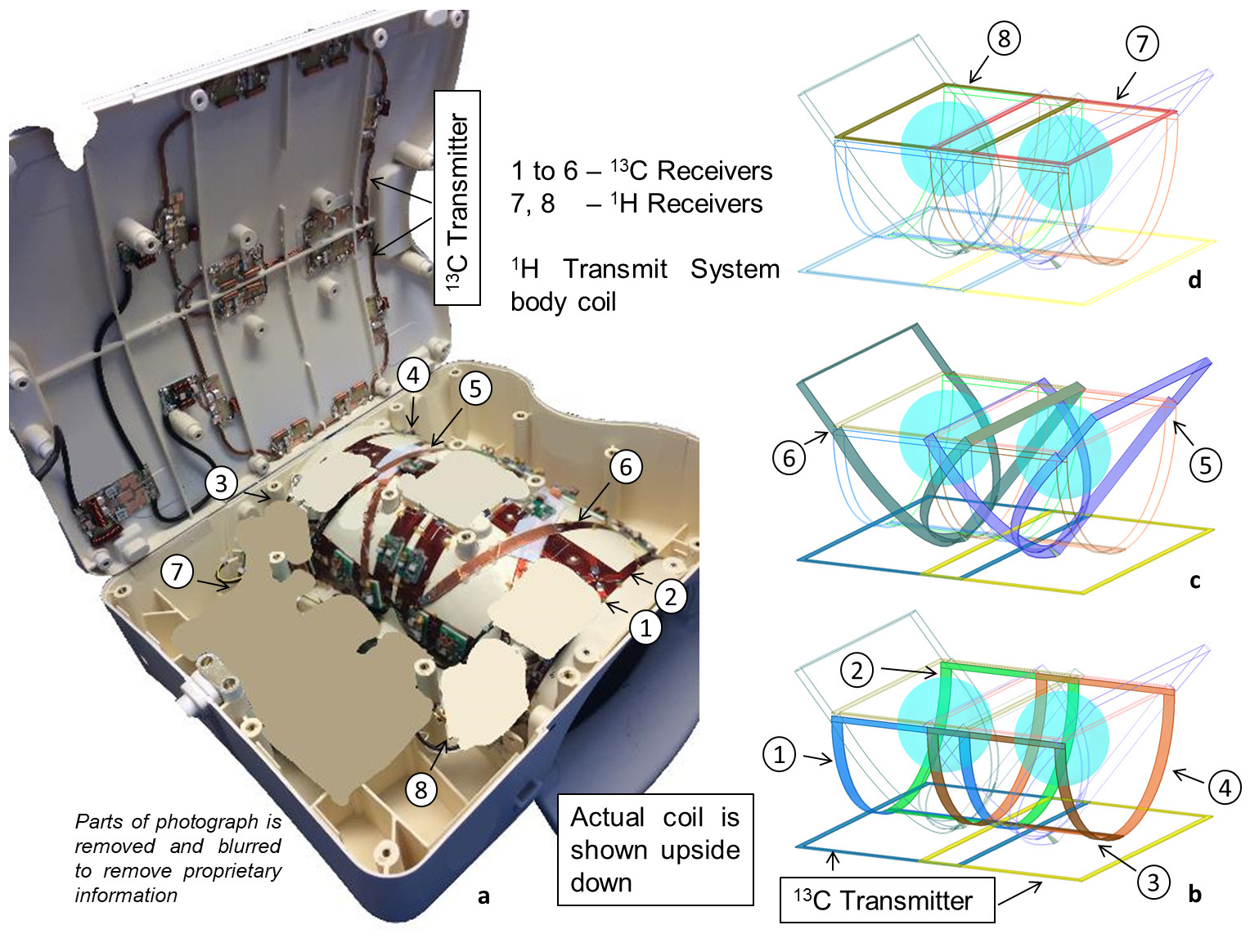
A former of an eight-channel breast coil for 1.5 T from a MR scanner manufacturer was repurposed to build the RF coil. The topology of the RF coil was modified from the original design to improvise the filling factor as shown in Figure 1. Of the eight 1H channels, 6 channels were tuned down to 13C at 3.0 T (32.35 MHz) and 2 channels were tuned up to 1H at 3.0 T (127.72 MHz), and low noise amplifiers were re-tuned to the respective frequencies. 1H traps were fitted to 13C channels and 13C traps were fitted to 1H traps as described earlier6. Channels 1 to 4 are mutually isolated using overlap, and isolated with 5 and 6 due to orthogonal polarization. Channels 5 and 6 are inductively decoupled with other. Channels 7 and 8 are isolated using overlap, and isolated with 13C channels using traps, and vice versa. Two RF coil loops for the 13C transmitter were built on the base plate of the former using hollow copper tubes, such that each loop is centred on each breast. Equal power was fed to the coils in-phase using a 2-way power divider. Both RF and MRI measurements in phantoms were performed to assess the RF coil performance.MR spectroscopy and imaging was performed on a GE MR750w system. To assess the performance of the RF coil for 13C NMR, spectroscopy was performed using a 13C-enriched bicarbonate (45 mL) phantom and chemical shift imaging (CSI) was performed in 3D using a 1.5 L cylinder and a 500 mL sphere filled with dimethyl silicone phantom. The spectroscopy parameters were; flip angle (FA) = 90°, TR = 2 s and 32 number of averages. The imaging parameters for 3D CSI were; axial plane, FA = 90°, TR = 2 s, field of view = 32 x 32 x 12 cm3 and matrix = 16 x16 x 6. Both spectroscopy and CSI were reconstructed with 10 Hz exponential apodization. 3D CS images were zero filled to 1 cm isotropic voxels. To assess the performance of the RF coil for 1H MRI, T1 weighted MR imaging was performed using water phantom doped with CuSO4. The imaging parameters were; FA = 10°, TE = 2.1 ms, TR = 3.5 ms, field of view = 42 x 43 x 30 cm3 and matrix = 300 x 300 x 60.
Results
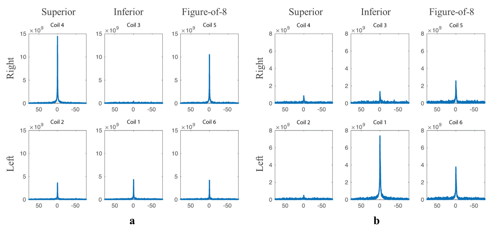
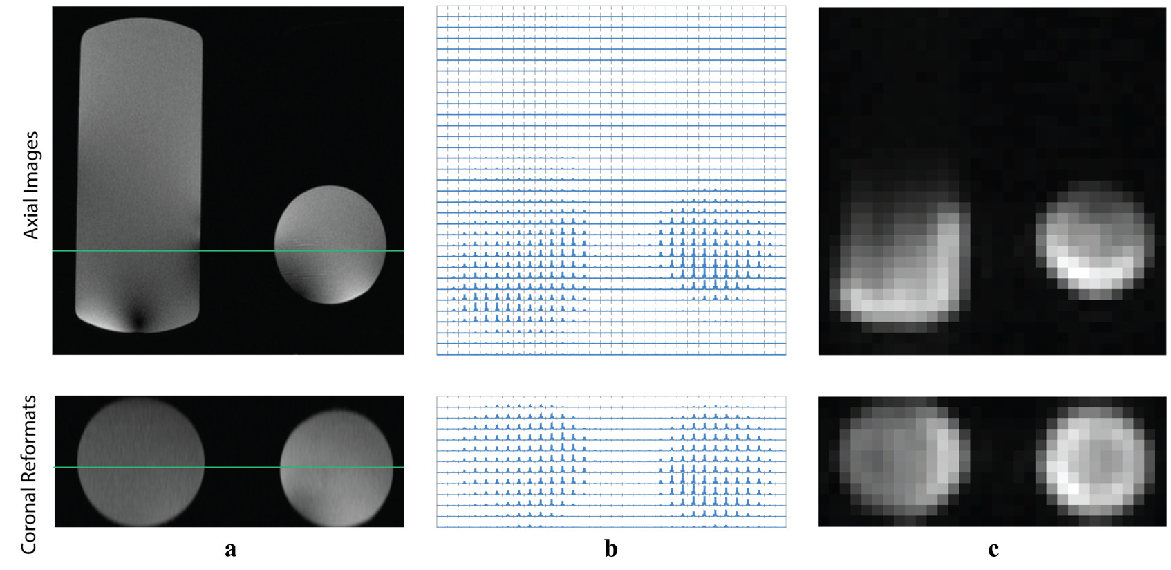
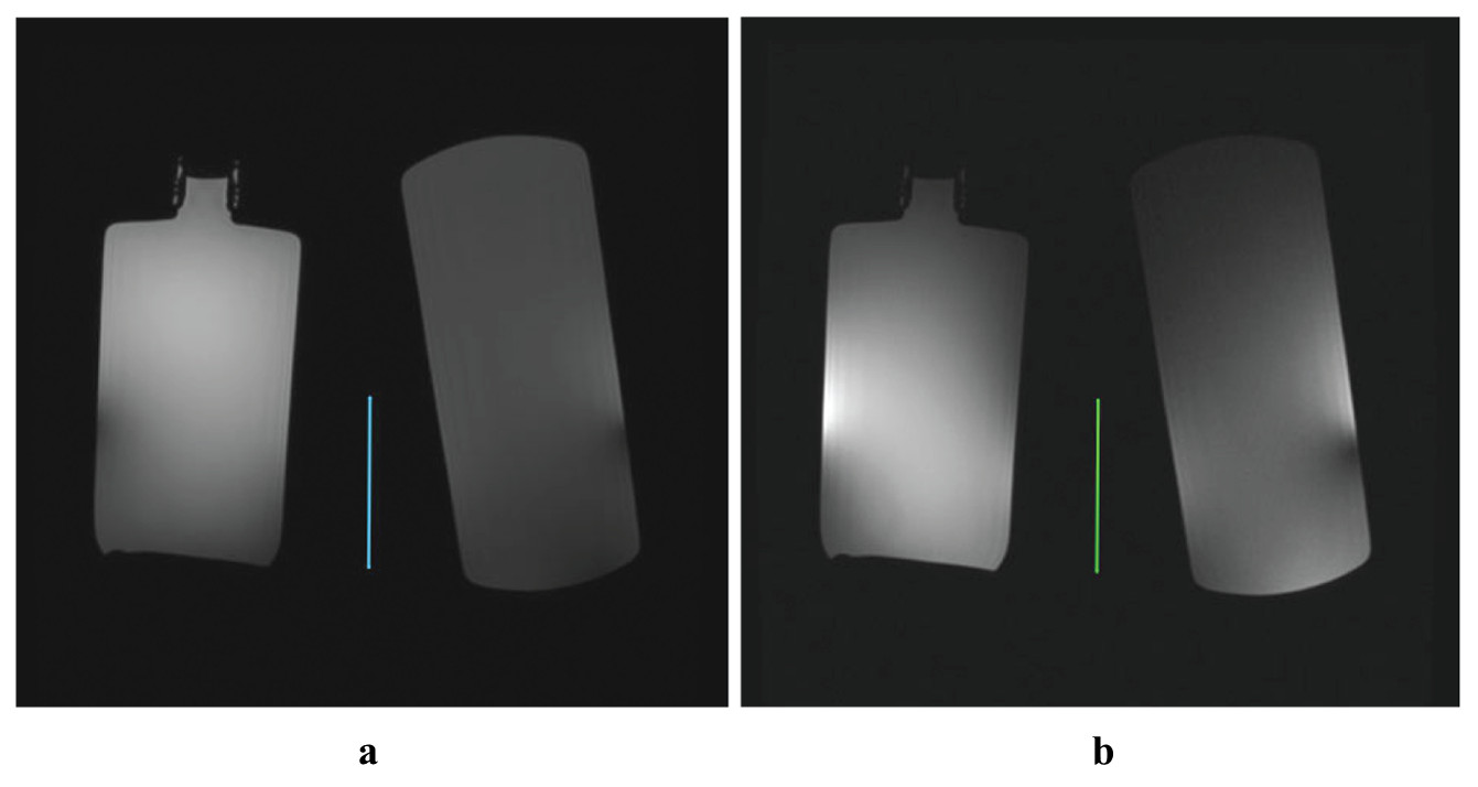
The worst case isolation of -16 dB was between channels 5 and 6. Isolation between 1H and 13C channels was -60 dB. The average unloaded and loaded Q factor for 13C elements were 230 and 80 respectively. 13C spectra obtained at the individual 6 channels of the RF coil are shown in Figure 2. Spatially resolved spectra and CS image obtained with 3D CSI are shown in Figure 3. 1H images obtained from the multinuclear 13C-1H breast RF coil and the system body coil for reference are show in Figure 4.Discussion
The RF coil channels for 13C benefited from improvised filling factor and 3D CS images were obtained with good image quality. For 1H channels, the design was limited by the physical form-fit of the repurposed former, where in the loop dimensions were unusually large considering the resonance frequency (127.72 MHz). Thus, to avoid very low capacitor values, the number of breakpoints to fit capacitors was drastically increased. Even then, some residual interaction of 1H channel with 1H traps on 13C channels was observed by examining the drop in unloaded Q factors when 1H traps were added, which may hinder sensitivity to some extent. Nevertheless, 1H images were obtained with good image quality. The in-vivo performance of this multinuclear 13C-1H breast RF coil in comparison to contemporary RF coil designs is yet to be established, and is the scope of this ongoing work.Conclusion
A design of a RF coil with 6 channels to receive 13C, 2 channels to receive 1H and with built in 13C transmitter is presented along with the demonstration of MR performance.Acknowledgements
This work was funded by the Engineering and Physical Sciences Research Council (EPSRC - EP/D070252/1), Medical Research Council (MRC - MR/M008894/1), National Institutes of Health (NIH R01 CA195476 and S10 OD016422; NIH/NCI Cancer Center Support Grant P30 CA008748) and the Peter Michael Foundation.
References
1. Wang ZJ, Ohliger MA, Larson PEZ, Gordon JW, Bok RA, Slater J, Villanueva-Meyer JE, Hess CP, Kurhanewicz J, Vigneron DB. Hyperpolarized 13C MRI: State of the Art and Future Directions. Radiology 2019;291(2):273-284.
2. Heuser L, Spratt JS, Polk Jr. HC. Growth rates of primary breast cancers. Cancer 1979;43(5):1888-1894.
3. Förnvik D, Lång K, Andersson I, Dustler M, Borgquist S, Timberg P. ESTIMATES OF BREAST CANCER GROWTH RATE FROM MAMMOGRAMS AND ITS RELATION TO TUMOUR CHARACTERISTICS. Radiation Protection Dosimetry 2016;169(1-4):151-157.
4. Kurhanewicz J, Vigneron DB, Ardenkjaer-Larsen JH, Bankson JA, Brindle K, Cunningham CH, Gallagher FA, Keshari KR, Kjaer A, Laustsen C, Mankoff DA, Merritt ME, Nelson SJ, Pauly JM, Lee P, Ronen S, Tyler DJ, Rajan SS, Spielman DM, Wald L, Zhang X, Malloy CR, Rizi R. Hyperpolarized (13)C MRI: Path to Clinical Translation in Oncology. Neoplasia (New York, NY) 2019;21(1):1-16.
5. Granlund KL, Morris EA, Vargas HA, Lyashchenko SK, DeNoble PJ, Sacchini VA, Sosa RA, Kennedy MA, Nicholson D, Guo YW, Chen AP, Tropp J, Hricak H, Keshari KA. First-in-woman study of in vivo breast cancer metabolism using hyperpolarized [1-13C] pyruvate Proc Intl Soc Mag Reson Med 24 P 3690 2016.
6. Rao M, Robb F, Wild JM. Dedicated receiver array coil for 1H lung imaging with same-breath acquisition of hyperpolarized 3He and 129Xe gas. Magnetic Resonance in Medicine 2015;74(1):291-299.
Figures