4100
Asymmetrical Body Birdcage Coil for Xenon-129 Imaging at 3T1Imaging Research Center, Cincinnati Children's Hospital Medical Center, Cincinnati, OH, United States, 2Center for Pulmonary Imaging Research, Cincinnati Children's Hospital Medical Center, Cincinnati, OH, United States, 3Philips Healthcare, Gainesville, FL, United States, 4Philips Healthcare, Best, Netherlands
Synopsis
Hyperpolarized 129Xe imaging is increasingly viewed as a viable tool for assessing lung structure and function. An asymmetrical whole-body xenon birdcage design for homogeneous transmit performance, previously evaluated through electromagnetic field simulations, was constructed. Coil homogeneity was evaluated with a high-pressure thermally polarized xenon Boltzmann cylinder phantom and hyperpolarized 129Xe gas. MR images of the phantom and the hyperpolarized gas confirmed homogeneous excitation.
Introduction
MR hyperpolarized 129Xe imaging has the potential to become a mainstream imaging technique for pulmonary disease and treatment assessment. Dedicated imaging coils with a homogeneous excitation volume over the lung region are required for exceptional xenon image quality. This is a challenge for birdcage coils not centered in the RF shield. Previously conducted electromagnetic B1+ homogeneity analysis of multiple birdcage designs1 identified an optimal asymmetrical xenon transmit/receive birdcage coil which was constructed for a Philips 3T Ingenia system. Imaging experiments to assess coil homogeneity were performed with a pressurized xenon gas Boltzmann cylinder and with a 6L bag containing a mixture of hyperpolarized 129Xe gas and nitrogen placed inside a hollow custom loader phantom2.Methods
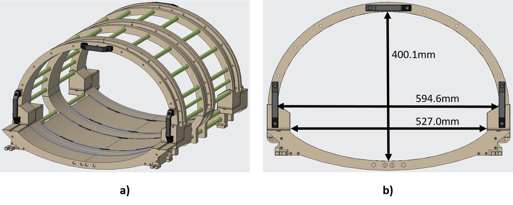
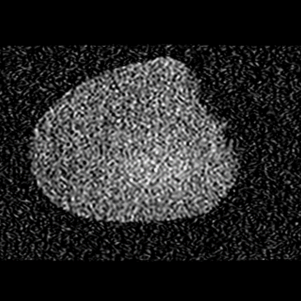
The birdcage coil was designed for a Philips 3T Ingenia 70cm bore system as a patient table mounted body coil with a detachable top half. The shell was constructed from four 3D printed endring sections, two middle sections, and two patient support inserts printed in ULTEMTM 9085 (Stratasys). Rungs were covered by 5/8” G10 tubing mounted between the individual ULTEMTM birdcage sections at rung locations. Shell thickness was limited to 1” along the ellipse dimensions to maximize patient space, Figure 1. Electrically, a high-pass birdcage design was implemented using the system’s proton body coil RF shield. Endrings were constructed from 1” wide and 1/8” thick copper bars and the legs from 1/4” copper tubing. Eleven rungs were distributed on the detachable top half and five rungs were distributed on the posterior table section of the coil. Electrical and mechanical connections of the birdcage's top and bottom sections were made in the lateral direction, versus traditional vertical direction, with Amphenol silver plated copper beryllium contacts. Xenon and proton baluns were placed on the I and Q drive ports to suppress common mode currents. Both ports were connected to a custom T/R switch, which connected to the MR system. Network measurements were performed with a Rohde & Schwarz ZNC3 network analyzer to assess tuning, matching, and coil Q for two adult males (84 kg and 65 kg). HFSS (ANSYS) was used for electromagnetic simulation of the asymmetrical birdcage loaded with a hollow torso loader phantom2 to visualize calculated B1+ distribution at different slice locations and for B1+ mean deviation inside a centered 25cm x 25cm x 17cm volume. Imaging experiments were performed with a hollow torso loader phantom containing a xenon high‐pressure high‐density polyethylene vessel cylinder filled with 61% Xe and 39% O2 at 11.6 bar2 and with a 6L bag containing a hyperpolarized 129Xe and nitrogen gas mixture. A gradient echo sequence (TR:750ms, TE:6.13ms, flip angle 74°, FOV:440 x 440mm, NSA:8, slice thickness: 100mm, recon voxel size 6.88mm x 6.88mm) was used for image acquisition of a single slice in the axial and coronal plane. An axial hyperpolarized 129Xe image was acquired with a gradient echo sequence (TR:9.5ms, TE:2.8ms, flip angle 9.2°, FOV:300 x 221.5mm, NSA:1, slice thickness: 15mm, recon voxel size 0.94mm x 0.94mm).Results
Loaded I and Q mode isolation was -44dB on a 84 kg male volunteer with a matching of S11 at -18dB and S22 at -13dB. A 65 kg male volunteer measured ‑23dB isolation, a matching of -23dB for S11, and -12dB for S22. Unloaded-to-loaded Q ratios for I and Q were greater than 2 for both volunteers. Electromagnetic simulation for the custom loader phantom showed a homogeneous B1+ distribution in all three planes (Figure 3). Analysis of the of simulated B1+ field distribution inside a centered rectangular box (25cm length, 25cm width, and 17cm height) showed 95% of the field was within 10% deviation of the mean. MR Images of the thermally polarized xenon Boltzmann phantom confirmed a homogeneous imaging profile, Figure 4. Imaging performed on a 6L bag containing a hyperpolarized 129Xe and nitrogen gas mixture further demonstrated the homogeneous imaging performance of the coil (Figure 5).Discussion and Conclusion
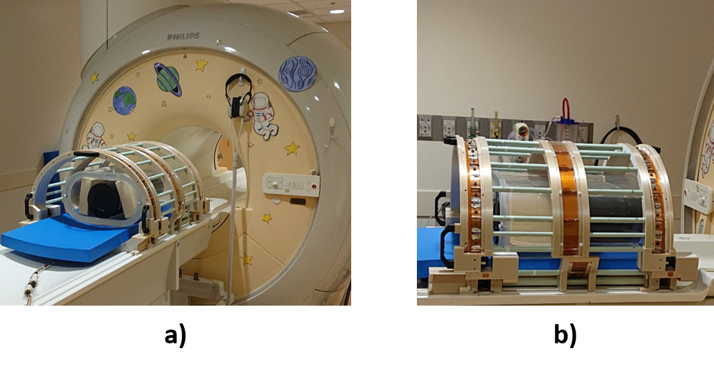
The detachable top half of the birdcage design allowed for obstruction free phantom and volunteer positioning. Connector technology and orientation eliminated the requirement for a dedicated locking feature of the two birdcage halves. The asymmetric design maximizes the available space for patients by conforming to the geometry of the MR patient table and bore wall. Mode isolation was similar to previously reported isolation of an elliptical birdcage for hyperpolarized 3He3 and better than reported for an asymmetric birdcage transmit coil for hyperpolarized 129Xe4. S11 and S22 were close to the values found for an asymmetric birdcage transmit coil for hyperpolarized 129Xe4. Electromagnetic simulation results of homogeneous excitation were confirmed with MR images on the xenon phantom and the hyperpolarized 129Xe gas bag image.Acknowledgements
No acknowledgement found.References
1. Loew W, Ireland C, Clevland Z, et al. Development of an Asymmetrical Birdcage Design towards Homogeneous Volume Excitation for Hyperpolarized Xenon-129 Lung Imaging at 3T. ISMRM 2018, #4408.
2. Bier E A, Nouls J C, Wang Z, et al. A thermally polarized 129Xe phantom for quality assurance in multi‐center hyperpolarized gas MRI studies. Magn Reson Med 82:1961–1968 (2019).
3. Zanche N D, Chhina N, Teh K, et al. Asymmetric Quadrature Split Birdcage Coil for Hyperpolarized 3He Lung MRI at 1.5T. Magn Reson Med 60:431–438 (2008).
4. Dregely I, Ruset I C, 2 Wiggins G, et al. 32-Channel Phased-Array Receive with Asymmetric Birdcage Transmit Coil for Hyperpolarized Xenon-129 Lung Imaging. Magn Reson Med 70:576–583 (2013).
Figures