4097
Evaluation of a Four-Ring Birdcage Head Coil for 1H Imaging and 13C Spectroscopy at 3 Tesla1NINDS, National Institutes of Health, Bethesda, MD, United States, 2NCI, National Institutes of Health, Bethesda, MD, United States, 3Lambda-Z Technologies, Baltimore, MD, United States, 4NCI, NIH, Bethesda, MD, United States
Synopsis
Dissolution DNP with Hyperpolarized 13C requires receive coils optimized for sensitivity and a transmit coil capable of delivering strong B1 fields for the carbon frequency. Here, a dual-tuned, four-ring birdcage head coil is described that can produce a transmit field of >90 microTesla and serve as a receive coil for an MRI system with few receivers. Hyperpolarized 13C spectra localized using 2D-CSI were obtained in 6 seconds from a human pancreatic tumor xenograph grown in the flank of a nude mouse. SNR of 13C spectra obtained from a tumor volume of <1 ml was suitable to measure the lactate/pyruvate ratio.
Introduction
Dissolution dynamic nuclear polarization (DNP) techniques1 have made it possible to acquire sensitive 13C metabolic images from prostate2, and brain3,4. The low gyromagnetic ratio of 13C requires strong transmit B1 fields to excite 13C spins. For head imaging, nested birdcage designs with 8 struts per layer are often used5. The four-ring birdcage, however, utilizes a single layer of conductors to provide superior axial B1 homogeneity at both frequencies6. Here, a 16-leg four-ring birdcage is described that provides excellent baseline performance for 1H imaging and 13C metabolic imaging at 3 Tesla. To provide a measure of the 13C sensitivity that might be obtained from a tumor in human brain, localized 13C spectra were acquired from a small (1 ml) human pancreatic tumor MiaPaCa-2 xenograft grown on the flank of the hind leg of a nude mouse.Methods
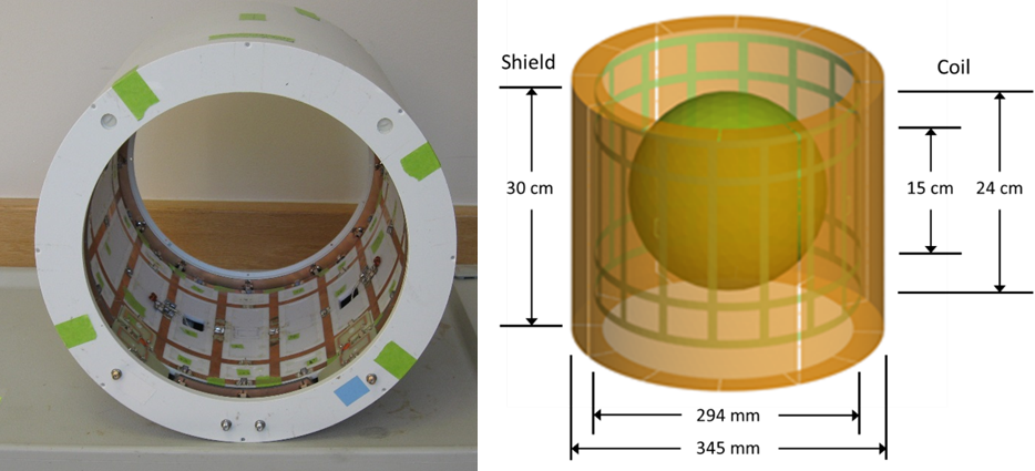
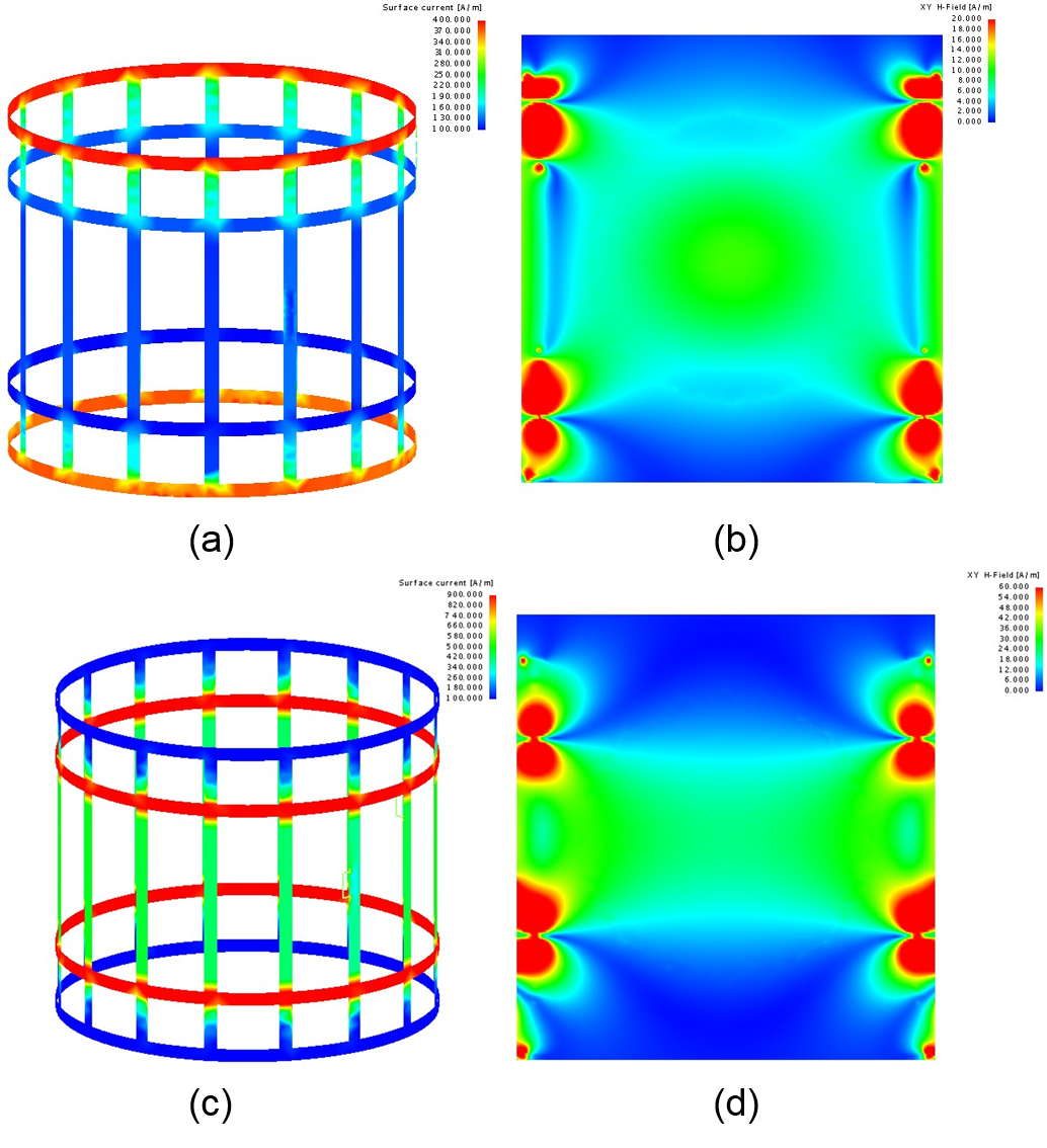
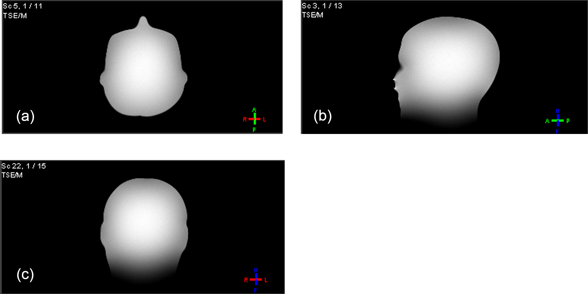
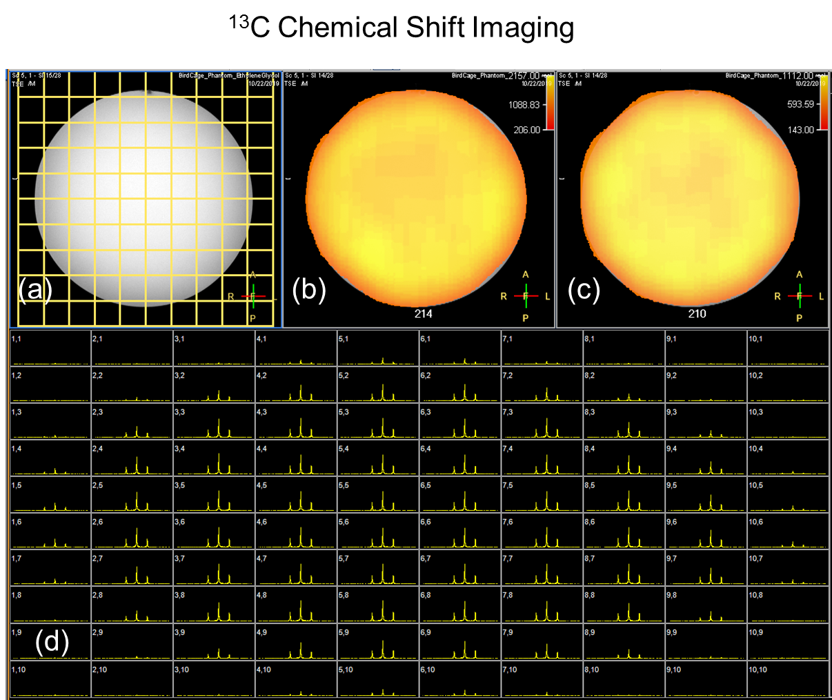
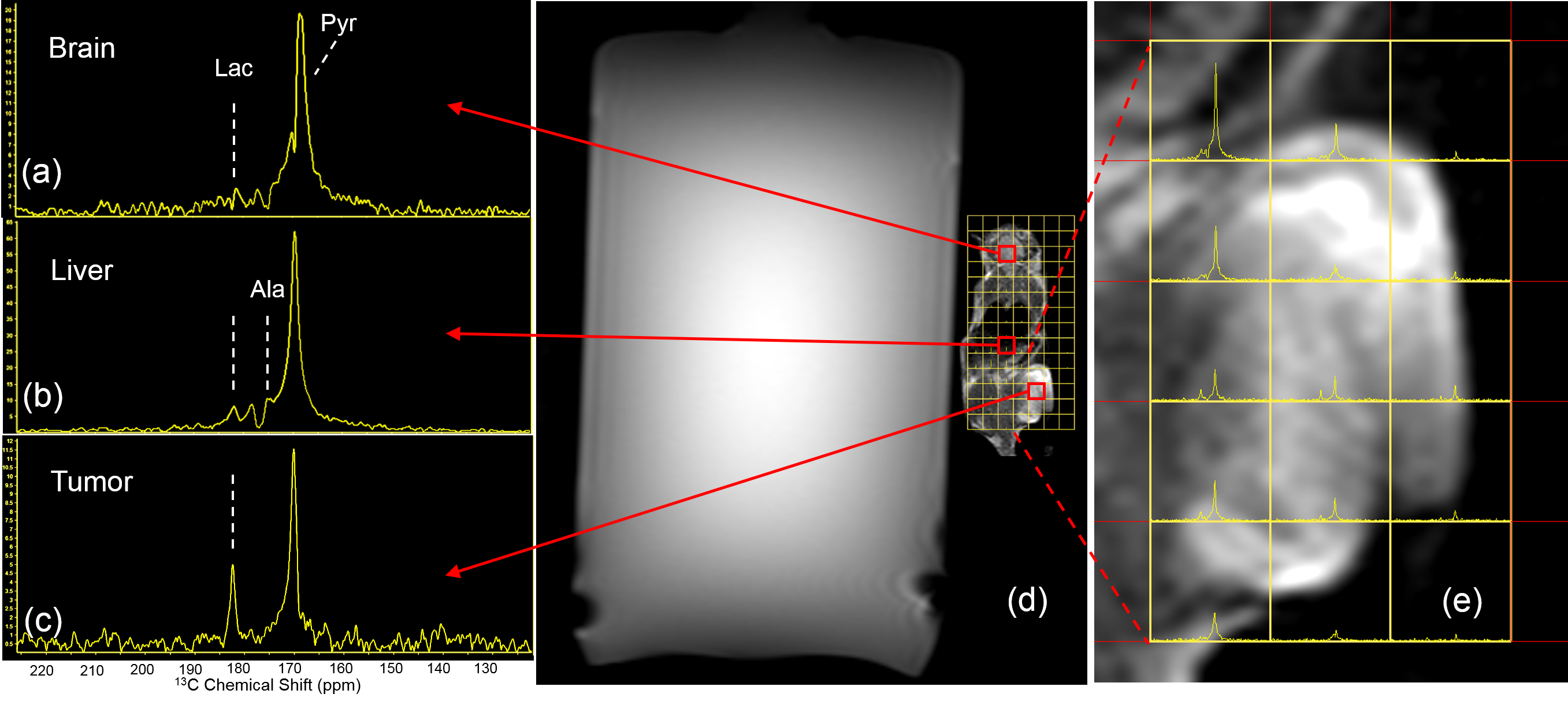
A dual-tuned, four-ring birdcage coil6, was designed as a transmit/receive coil for 1H imaging and 13C spectroscopy of the head. The coil, shown in Fig. 1a, was constructed with 16 legs, mounted concentrically inside an RF shield, and configured to operate in quadrature mode at both frequencies. The dimensions and ring spacings for the coil, shown in Fig. 1b, were chosen to be proportionally the same as for a dual tuned body coil reported earlier7. Four saddle-type inductive couplers were mounted over outer meshes of the coil as elements of a four-port coupling network for the 1H frequency. For the 13C frequency, two quadrature feed cables were routed over the RF shield to matching points on the inner legs of the coil. Traps for the 1H frequency were inserted in series with these connections to improve isolation. Tuning of the coil was performed on the bench using a head-shaped phantom (Phantom Labs, Niskayuna, NY) filled with PVP and saline (ε = 52, σ = 0.55). The RF currents and fields of the coil were simulated at both frequencies using FEKO software (Altair Hyperworks, Troy, MI). For the 1H field simulations, Thevenin sources were connected across four outer mesh capacitors. In Fig. 3, gradient echo 1H images were acquired from the head phantom on a Philips Ingenia Elition 3T scanner, and 1H sensitivity was compared with that of the standard head coil. To evaluate 13C sensitivity, 2-D CSI spectra were acquired from an 18 cm spherical phantom filled with 100% ethylene glycol and processed using R5.6.0 software on the scanner. Finally, localized hyperpolarized 13C spectra were acquired from a pancreatic tumor xenograft grown in the flank of a nude mouse. Bolus 96 mM [1-13C]pyruvate, which was generated by SPINLAB (GE Healthcare, Milwaukee, Wisconsin), was intravenously injected to the animal 10 seconds after dissolution, and 2-D localized CSI spectra were acquired from the entire mouse 30 sec after injection.Results
Tuning of the four-ring birdcage for 1H and 13C was straight-forward, since none of the high-order modes of the birdcage structures were overlapping. 1H port to 13C port isolations were greater than -45 dB for both frequencies. RF field simulations for each frequency are shown in Figs. 2b and 2d for coronal views through the coil. In sagittal views,1H images of the head phantom show good uniformity, with image intensity dominated by the dielectric properties of the phantom. 1H sensitivities measured at the center of a silicon oil phantom were 169 for the four-ring birdcage and 173 for the standard head. For the 13C frequency, simulations in Fig. 2b showed good coverage over the brain. Bench measurements of a loaded coil using a calibrated loop yielded a B1 of 1.68 µT/ Watt1/2. The maximum available transmit power for 13C was 3150 Watts, yielding a maximum B1 for the coil of 94 µT. This was confirmed with tip angle measurements of the phantom. In Fig. 4, 2-D localized 13C spectra acquired from the 13C phantom show baseline resolved triplets from ethylene glycol. Spectroscopic images in from the center and side peaks showed good uniformity, with image intensities stronger near the coil. Nude mice were injected with hyperpolarized [1-13C]pyruvate, and 2-D CSI spectra were acquired in 6 seconds in a 7 x 14 array positioned over the animal (Fig. 5). Spectra shown were obtained from 5mm x 5mm x 15mm voxels from the brain, liver, and tumor located in the hind leg.Discussion
The four-ring birdcage provides excellent coverage for the head with minor loss in 1H sensitivity as compared with a single-tuned coil for an MRI system. The maximum transmit B1 field achieved for the 13C frequency was 94 µT, more than double that of a “clam shell” coil provided with the 13C accessory. Because the 13C accessory provides only six receiver channels, a quadrature coil provides near optimal sensitivity as compared with a limited array. Here, the four-ring birdcage provided enough sensitivity from a <0.5 ml tumor voxel to resolve and quantify the lactate-to-pyruvate ratio.Conclusion
The four-ring birdcage provides excellent 1H image uniformity and excellent transmit B1 capability for hyperpolarized 13C experiments. 13C sensitivity and coverage are adequate for metabolic imaging of the human brain. The four-ring birdcage provides near optimal performance as a quadrature coil and provides a competitive baseline with which to compare head arrays3,4.Acknowledgements
This research was supported (in part) by the Intramural Research Programs of the NIH, NINDS and NCI.References
1. Ardenkjaer-Larsen J-H, Fridlund B, Gram A, et al. Increase in signal-to-noise ratio of >10,000 times in liquid-state NMR. PNAS 2003;100(18):10158.
2. Nelson S, Kurhanewicz J, Vigneron D, et al. Metabolic Imaging of Patients with Prostate Cancer Using Hyperpolarized [1-13C]Pyruvate. Sci Translational Med 2013;5(198):1.
3. Mareyam A, Carvajal L, Xu D, et al. 31-Channel brain array for hyperpolarized 13C imaging at 3T. Proc. ISMRM 2017;25:1225.
4. Ma J, Hashoian R, Sun C, et al. Development of 1H/13C RF Head Coil for Hyperpolarized 13C Imaging of Human Brain. Proc. ISMRM 2019;27:568.
5. Fitzsimmons J, Beck B, Brooker R. Double Resonant Quadrature Birdcage. Magn Reson Med;1993;30:107-114.
6. Murphy-Boesch J, Srinivasan R, Carvajal L, et al. Two Configurations of the Four-Ring Birdcage Coil for 1H Imaging and 1H -Decoupled 31P Spectroscopy of the Human Head. J Magn Reson 1994;103B:103-114.
7. Boskamp E, Xie Z, Taracila V, et al. A Dual-Tuned 70 cm Whole-Body Resonator for 13C and Proton MRI/MRS at 3T. Proc. ISMRM 2018;26:1714.
Figures