4080
Novel ceramic-based resonators for bilateral breast imaging at 3T1Faculty of Physics and Engineering, ITMO University, Saint-Petersburg, Russian Federation, 2Research and Practical Clinical Center of Diagnostics and Telemedicine Technologies of the Moscow Heathcare Department, Moscow, Russian Federation
Synopsis
We propose a concept for targeted bilateral breast imaging. A practical demonstration of the concept features a pair of hollow dielectric cylindrical resonators based on a composite material with very high permittivity. The resonators are electromagnetically coupled to the body birdcage coil and each other. Electromagnetic simulations demonstrated that symmetric B1+ field mode, inherent in interacting resonators, can be efficiently excited at 123 MHz via the body birdcage coil. Simulations with realistic voxel models let us estimate transmit efficiency and radiofrequency safety of the proposed device and demonstrated that the concept promising for clinical applications.
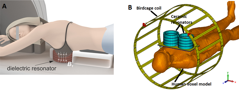
Here we describe and demonstrate through electromagnetic simulations, a concept of targeted bilateral breast MRI, as a way to increase specificity of breast imaging for screening purposes. The idea is based on a local redistribution and passive focusing of the radiofrequency (RF) magnetic flux of the birdcage coil (BC) using electromagnetic coupling with subwavelength dielectric resonator surrounding the target area (Figure 1A). Proof-of-concept unilateral targeted breast imaging was numerically and experimentally approved in our previous work.1 In this work we introduced two coupled resonators for bilateral imaging, moved to appropriate size of hollow ceramic cylinders and estimated RF safety with realistic voxelised models, thus making a step towards clinical application.
Methods
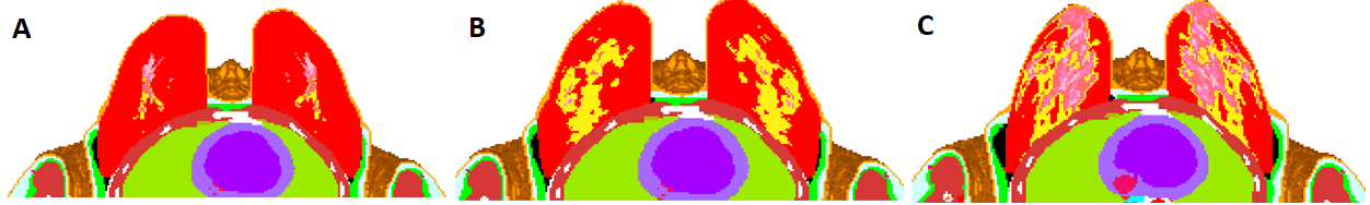
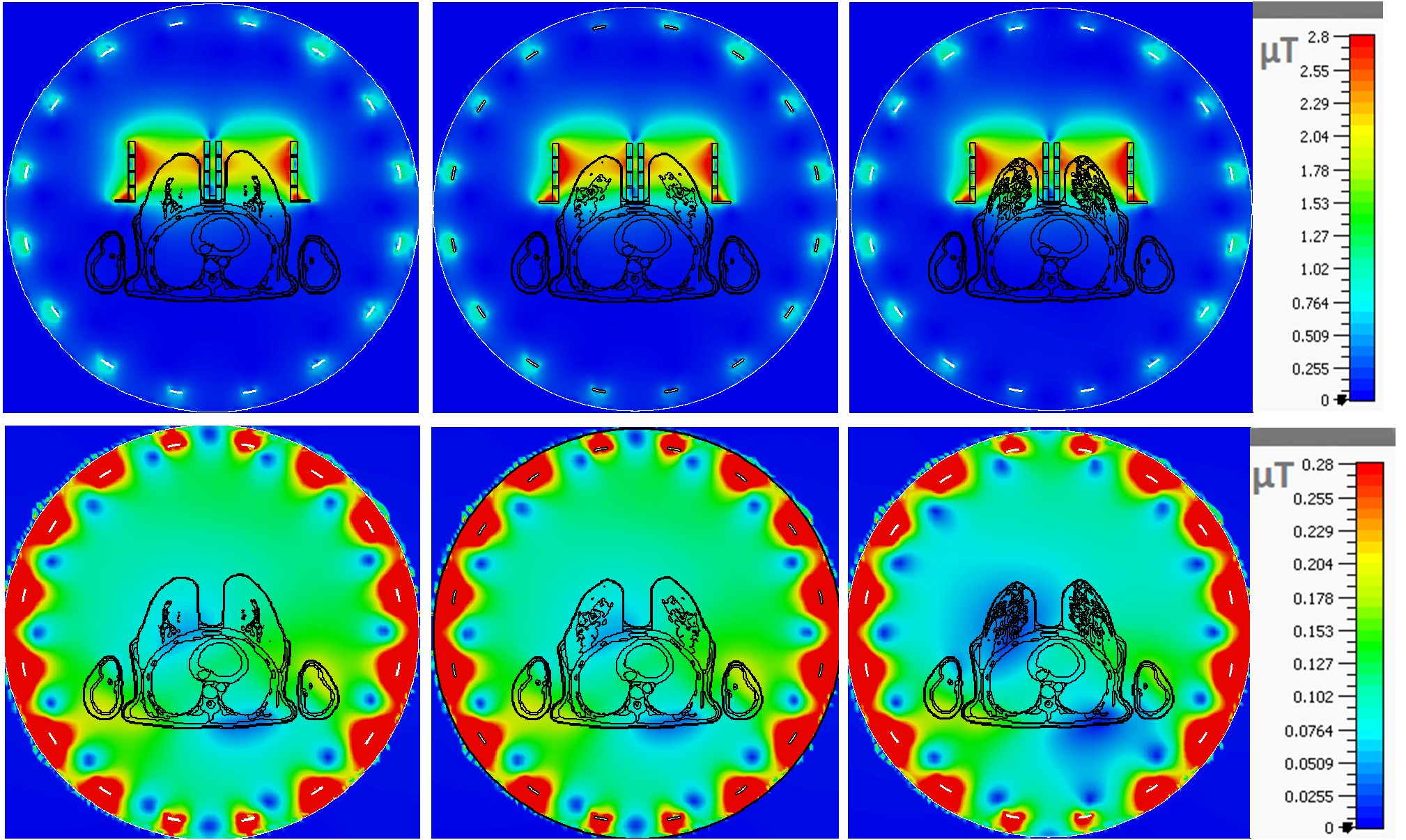
A proposed concept was based on a passive focusing of B1+ field of a body birdcage coil using a high permittivity dielectric resonators.2,3 Each resonator was constructed of four ceramic rings with relative permittivity ε≈900 and tanδ ≈0.0004 (at 1 MHz) and the following dimensions: inner diameter 128 mm, outer diameter 150 mm, the height of each ring 25 mm. Distance between the centres of the resonators was 160 mm. The placement of two cylindrical resonators next to each other causes magnetic and electrical field mode splitting due to the electromagnetic coupling. In other words, resonator’s TE01δ-mode4 was split into asymmetrical (lower frequency) and symmetrical (higher frequency) modes. The frequency difference between asymmetrical and symmetrical modes was about 10 MHz. As far as distribution of B1+ field in left and right breasts should be the same, it is necessary to excite symmetrical magnetic field mode of the coupled resonators. The frequency of the symmetrical mode was tuned to 123 MHz (3T MRI) by changing the spacing between the ceramic rings. We have used several thin rings made of plexiglass with ε~3.5 and σ=0.02 S/m as spacers, which had the same inner and outer diameters as the ceramic rings. To preserve the efficiency of the ceramic resonators with proximity to the chest a thin metallic plate of 35 μm-thick copper with two holes (with the same diameter as the outer diameter of ceramic rings) was added. All numerical simulations were performed in CST Microwave Studio 2017. Mode splitting was investigated as a function of the distance between the resonators. Here we used a loop coil placed at the centre over one of the resonators to excite TE01δ-mode (Figure 3A, B). Then electromagnetic modelling of the resonators was performed using voxelized female human body models placed in a standard birdcage coil (Figure 1B). We considered three voxel models5 with different ratio between glandular and fatty tissue and its distribution over the breasts: Phantom #1 from ACR Class 1, #3 from ACR Class 2 and #1 from ACR Class 4 (Figure 2). Breast shape and position of the models corresponds to the physiological configuration during conventional breast imaging. For a reference, the birdcage coil was simulated with the same body models at the same position, but without the resonators. The B1+ field distributions were normalized to 1W of total accepted power. The SARav.10g distributions were normalized to the same B1+ averaged in the central XY slice in the breast area. The effect of the resonators on the birdcage coil transmit performance was evaluated by comparison of the ratio $$$\frac{|B1+RMS |}{\sqrt{psSARav.10g}}$$$, denoted by RF safety , in the presence of the resonators to the ratio with the BC alone, where the field value was spatially averaged over the targeted area (RMS=root mean square), and psSARav.10g is the 10-g-averaged peak spatial SAR.
Results
Figure 3 demonstrates mode splitting for the different distance between the resonators: 16, 18, 20 cm. The closer the resonators are, the stronger is coupling between them, and the bigger is frequency gap between the modes and the lower is the quality factor of the symmetrical mode. Consequently, ceramic resonators efficiency became 15% lower for 20 cm case than for 16 cm. Figure 4 show that the ceramic resonators, being tuned to the symmetrical mode, coupled effectively to the birdcage coil and focused its B1+ field in the breasts’ areas. Figures 5 demonstrate peak spatial SAR value decrease. RF safety grew by 6.6 fold to reference case for mostly fatty breast (Figure 2A), by 11.7 fold for scattered fibro glandular breast (Figure 2B), and by 14.1 fold in case of very dense breast (Figure 2C).
Discussion and conclusions
A couple of high permittivity ceramic resonators can be used for targeted bilateral breast MRI at 3T. The resonators effectively redistribute the electromagnetic field of the body birdcage coil and localise it around the areas of interest that boosts both transmit efficiency and RF safety of the breast imaging. 16 cm distance between the centres of the resonators was chosen considering average anatomical dimensions, and it is not optimal regarding the symmetrical mode quality factor. Nevertheless, such configuration already gives significant benefits in terms of transmit efficiency and RF safety (up to 23.8 and 14.1 fold respectively in case of the very dense breast). Further investigations (for example, B1+ field inhomogeneity improvement over the region of interest) of the resonator and MRI experiments with volunteers are ongoing.
Acknowledgements
This work was supported by the grant of the Russian Science Foundation (Grant No. 18-75-10088).
The authors are grateful to Dr Arthur W. Magill for providing realistic voxel models.
References
1. Shchelokova A, et al. Dielectric resonator for targeted breast MRI at 3T. Proc. Intl. Soc. Mag. Reson. Med. 2019; 27, 0435.
2. Mett R R et al. Dielectric microwave resonators in TE011 cavities for electron paramagnetic resonance spectroscopy. Rev Sci Instrum. 2008;79:094702.
3.Webb A. Dielectric materials in magnetic resonance. Concept Magn Reson A. 2011 ;38A: 148-184.
4. Webb A. Cavity- and waveguide-resonators in electron paramagnetic resonance, nuclear magnetic resonance, and magnetic resonance imaging. Prog Nucl Mag ResSp. 2014;83:1-20.
5. The UWCEM Numerical Breast Phantom Repository http://uwcem.ece.wisc.edu/phantomRepository.html
6. Tissue properties https://itis.swiss/virtual-population/tissue-properties/overview/
Figures