4047
Stacked dipole arrays for 31P and 1H MRI in the body at 7T1Radiology, UMC Utrecht, Utrecht, Netherlands, 2UMC Utrecht, Utrecht, Netherlands, 3TeslaDC, Zaltbommel, Netherlands, 4Oxford Centre for Magnetic Resonance Research, University of Oxford, Oxford, United Kingdom, 5Dept of Clinical Neurosciences, University of Cambridge, Cambridge, United Kingdom, 6Biomedical Engineering, Eindhoven University of Technology, Utrecht, Netherlands
Synopsis
While an RF birdcage integrated behind the bore-liner of a 7T MRI can provide relatively uniform 31P excitation, it requires substantial system integration activities. Here we present an alternative approach using an array of stacked dipole antennas where one is tuned to 1H and the other to 31P. The setup is significantly easier to configure and while 31P B1+ uniformity is somewhat compromised, B1+ is more efficient when compared to an integrated birdcage.
INTRODUCTION
Metabolic imaging using X nuclei is an attractive technique for it provides an unique insight into cellular metabolism, is not hindered by the three orders of magnitude higher water and lipid signals and is less sensitive to B0 and B1 non-uniformities. While ideal X-nuclei transmit coils would, similar to 1H transmit coils, be integrated behind the bore liner of the MRI system to maximize patient comfort and provide uniform excitations, it comes at a substantial integration effort, sometimes neither supported by the MRI vendor nor the institute. Here we present an alternative approach for a 31P and 1H setup that maintains patient comfort and can provide good B1+ throughout the human body at 7T. We show by simulations and bench top measurements that the B1+ fields of dipoles tuned for 1H do not affect the fields of dipoles tuned for 31P so that they can be stacked together. When used as an 8 channel transceiver array, so far without stacking, good MRI performance is demonstrated when operated at the frequencies of 1H (demonstrated at 7T) and close to 31P (demonstrated by 1H MRI 3T).METHODS
A 30cm fractionated dipole was tuned to 298MHz (1H at 7T). Similarly, another 30cm dipole was tuned to 120MHz (31P at 7T) using continuous meandering. Efficiency was measured on a large saline filled phantom with 2cm separation between the phantom and closest antenna. The second antenna was placed with 1cm spacer on top of the other antenna. A cylindrical hole through the phantom was used to hold a calibrated pickup probe (fig1). B1+ and SAR simulations were performed using Sim4Life on Duke and S11 and S12 values were measured with the elements stacked, and for each separately. 1H and 31P ceramic cable traps (one of each; 1H closest to feed port) were placed on each cable to suppress shield currents. The efficiency (S12) was compared to an integrated 31P quadrature birdcage coil. To illustrate the potential in vivo performance of the dipole array at 31P (120MHz), an 8 channel dipole array was constructed and tuned for 3T 1H MRI (128MHz) and tuned for 1H MRI at 7T upon which MRI was obtained from a healthy volunteer.RESULTS
B1+ and SAR simulations did not reveal any field variations due to stacking at either the 31P or 1H frequency (Fig 2). Stacking order made no difference. For the benchtop measurements, the 31P element was positioned closest to the phantom. The S11 on phantom remained good (-17dB to -20dB) for 1H when comparing single element to the stacked element respectively, and S12 remained identical. For 31P, the S11 dropped from -15dB to -6dB at 120MHz, which resulted in a small reduction in S12 of 2dB when comparing the single element to the stacked element respectively. Here, the presence of the 1H element shifted the matched condition (-18dB) of the 31P element to 131MHz. The S12 of the single tuned 31P element was 2dB higher when compared to the S12 of a single port of the birdcage with the same pickup probe placed in the isocenter. MRI (in figure 3) that was obtained with the 8 channel dipole array reflects good uniformity in the body when driven close to the 31P frequency and fair uniformity when driven at the 1H frequency for 7T.DISCUSSION
A dipole array can be a good candidate for providing a relatively uniform B1 field, not only for 1H but also for 31P. While simulations show negligible effect of stacking the antennas, bench top measurements did show a load dependence resulting in a subtle effect on the matching conditions, which could be counteracted by adapting the matching network to regain the subtle loss in efficiency (S12). In contrast to double tuned setups that often compromise performance for one or both nuclei, here we do not observe the compromise. In comparison to a 31P birdcage body coil, the 31P dipole array has a 1.7-fold more favourable B1 and more than 1.3-fold increased SAR efficiency (below 4.3μT/ for the birdcage vs 5.7μT/with B1 normalised per square-root peak local SAR based on Duke simulations). However, the dipole array does require B1 shimming for ensuring uniform B1.CONCLUSION
An array of two stacked dipole antennas (one for 1H, one for 31P) can perform equally well as the corresponding arrays of 1H or 31P alone. The 31P antennas are more efficient than birdcages and therefore may be an attractive alternative to integrate X-nuclei MRI in traditional 1H RF coil arrays.Acknowledgements
This work was supported by: European H2020-FETOPEN: NICI
C. T. Rodgers is funded by a Sir Henry Dale Fellowship from the Wellcome Trust and the Royal Society [098436/Z/12/B].
References
- Sophie A. Kurk, Bart R. Steensma, et al.
Feasibility of 7-T fluorine magnetic resonance spectroscopic imaging (19F MRSI)for TAS-102 metabolite detection in the liver of patients with metastatic colorectal cancer. European Radiology Experimental. 2018; 2:16
- Alexander J.E. Raaijmakers,et al.
Fractionated Dipole Antenna: A New Antenna for Body Imaging at 7 Tesla. Magnetic Resonance in Medicine. 2016; 75:1366–1374
- Aidin Ali Haghnejad, Mark Gosselink, et al.
Fixed-phase prostate imaging with a 8-channel transmit/receive dipole antenna array on a conventional 3T system. Proceedings of the ISMRM 26th Annual Meeting 2018, # 1704
- Aidin Ali Haghnejad, Shaihan J. Malik, et al.
Transceive surface array of dipole antennas for multi-transmit imaging at 3T. Proceedings of the ISMRM 24th Annual Meeting 2018, # 3174
Figures
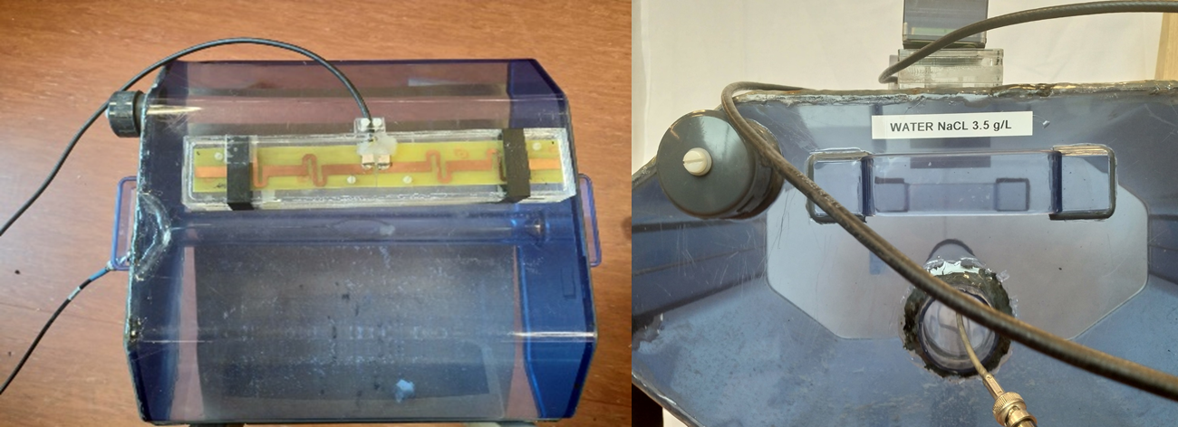
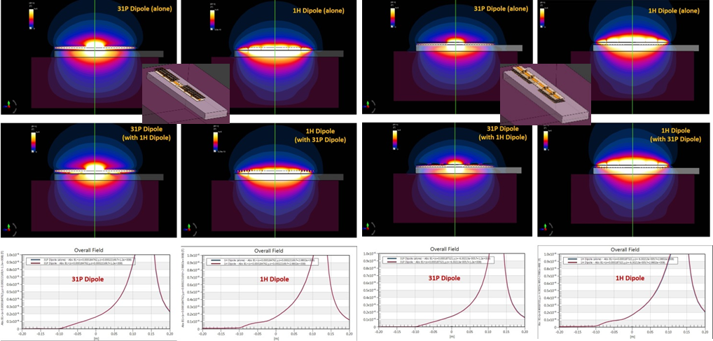
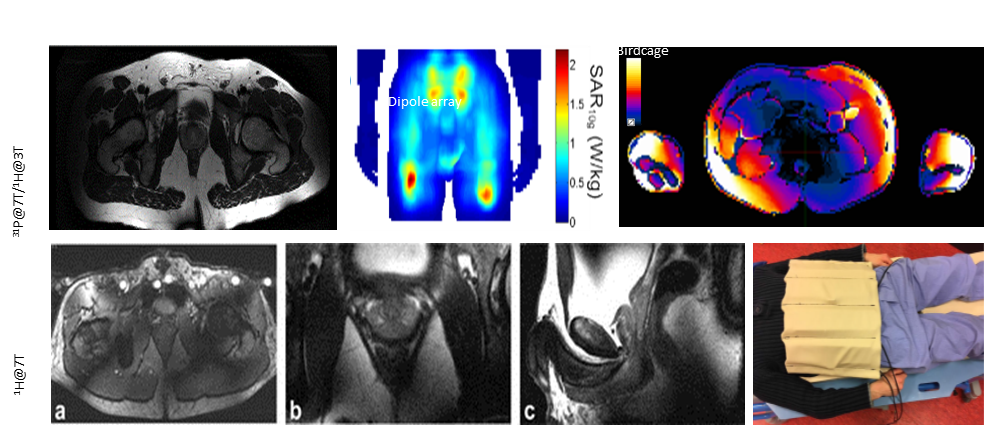
Figure 3: From left to right;
Top: MRI of prostate in volunteer using 8 channel 31P dipole array (tested by performing 1H-MRI at 3T at practically the same frequency), SAR simulation of 31P dipole array with 8 elements and, SAR simulation of 31P birdcage
Bottom: MRI of prostate in volunteer using 8 channel 1H dipole array in (a) transverse (b) coronal (c) sagittal planes and setup