4040
Evaluation of signal-to-noise ratio of a size-adaptable 16-channel head array
Kohjiro Iwasawa1, Yosuke Otake2, Kazuyuki Kato2, Hideta Habara2, Masayoshi Dohata2, Hisaaki Ochi1, and Toru Shirai1
1Research & Development Group, Hitachi, Ltd., Tokyo, Japan, 2Healthcare Business Unit, Hitachi, Ltd., Tokyo, Japan
1Research & Development Group, Hitachi, Ltd., Tokyo, Japan, 2Healthcare Business Unit, Hitachi, Ltd., Tokyo, Japan
Synopsis
In order to increase both coil setting efficiency and coil sensitivity, a size-adaptable tight-fit 16ch head array which enables one-action-fixation was investigated at 3 T. The anterior part of the head array is a flexible coil, and can fit to the patient when the fixation belt is tightened. SNR increased by 1.2-1.4 times across 1-99 percentile adult head sized phantoms against a conventional 15ch rigid head coil. SNR improvement could also be seen in vivo with a healthy volunteer.
Introduction
In order to increase head examination throughput, RF receiver coils are required to increase both coil sensitivity and coil setting efficiency. However, conventional rigid head coils have issues: (1) Unoptimized sensitivity due to distance between the coil and the patient’s head. (2) Takes time and effort for fixation of the patient (e.g. squash in sponges between the coil and the head to restrict motion). To address these issues for various-sized patients, size-adaptability is a key technology.1-9 In this study, we propose a size-adaptable tight-fit head array that closes the distance of the coil and head with simplified head fixation, in order to increase both coil sensitivity and coil setting efficiency. We made a 16-channel (ch) prototype and evaluated the signal-to-noise-ratio (SNR) of images acquired with a 3 T scanner.Methods
Size-adaptable tight-fit head arrayFigure 1 shows the coil setting procedure of a size-adaptable tight-fit head array. The anterior part of the head array is a flexible coil, and can fit to the patient when the fixation belt is tightened. At the same time of fitting the anterior coil, the head is firmly fixed by the fixation belt above the forehead and the fixation panels closing in from both sides. This one-action fixation enables shorter coil setting time without compromising fixation firmness. Figure 2a shows a 16ch prototype of a size-adaptable tight-fit head array. Size adapts by the position of the anterior coil (coil elements are fixed-length). Enhanced preamp decoupling via intentionally small matching capacitance9 was applied to minimize SNR degradation due to displacement of the anterior coil.
SNR evaluation
SNR of images acquired on a 3 T scanner with the prototype was compared to that of a conventional 15ch rigid head coil. SNR was calculated with optimum reconstruction accounting noise correlations.10
Phantom study (Fig. 2b)
Phantoms of three sizes were investigated. The perimeters of the phantoms ranged from 1 to 99 percentile adults (514, 591, and 619 mm respectively). Scan parameters were as follows: 2D GrE, TE/TR = 30/685 ms, BW 38.3 kHz, FA 90 deg., thickness 1 mm, FOV 256×256 mm2, Matrix 128×128. SNR was compared at two regions of interest; (1) Whole brain region: 8 voxels shrunk from the edge, (2) Center region: 30 mm diameter at the center of the middle slice (red circles in Fig. 3).
Volunteer study (Fig. 2c)
A healthy male volunteer was scanned. FLAIR images were compared where SNR can be comparably low among routine images. Scan parameters were as follows: 2D FIR, TE/TR = 132/9000 ms, BW 70 kHz, FA 90 deg., thickness 5 mm, echo factor 26, NSA 1, RAPID 1.0, FOV 230×230 mm2, Matrix 352×256. For post-processing, sensitivity correction was applied but image filters were not applied. Data were obtained according to the standards of internal review board on Research & Development group, Hitachi, Ltd.
Results
Figure 3 shows SNR maps of phantoms of three sizes. Results show that SNR improved especially in the anterior region of the small sized phantom. This is due to the distance between the coil and subject closing in compared to the large distance for a fixed-size rigid coil. The SNR in the right and left regions also improved due to the anterior coil being flexible and conform to the shape of the subject from the one-action fixation. SNR increased by 1.2-1.4 times in the whole brain region, and by 1.1-1.3 times in the center region for phantoms ranging across adult head sizes (Fig. 4). SNR improvement could also be seen in FLAIR images of a healthy volunteer (yellow arrows in Fig. 5).Discussion
Enhanced preamp decoupling via intentionally small matching capacitance9 lead to both close fit for higher SNR and decoupling enhancement. Further investigation is needed for magnetic field strength below 3 T, where matching capacitance tends to enlarge due to smaller sample loss.Conclusion
To increase both coil setting efficiency and coil sensitivity, a size-adaptable 16ch head array which enables one-action-fixation was investigated at 3 T. SNR increased by 1.2-1.4 times across 1-99 percentile adult head sized phantoms against a conventional 15ch rigid head coil.Acknowledgements
No acknowledgement found.References
- J. A. Nordmeyer-Massner, et al., “Stretchable Coil Arrays: Application to Knee Imaging,” Magn. Reson. Med., Vol. 67, pp. 872-879, (2012).
- B. Wu, et al., “Flexible Transceiver Array for Ultrahigh Field Human MR Imaging,” Magn. Reson. Med., Vol. 68, pp. 1332-1338, (2012).
- V. Taracila, et al., “Adaptive head array,” Proc. ISMRM, pp.4883, (2014).
- G. C. Wiggins, et al., “Size-adaptable “Trellis” receive array concept for knee imaging,” Proc. ISMRM, pp.0493, (2016).
- P. Rossman, et al., “Characterization of a new ultra-flexible, low profile RF receive coil technology,” Proc. ISMRM, pp.0763, (2017).
- N. L. Rios, et al., “Size-adaptable 13-channel receive array for brain MRI in human neonates at 3 T,” NMR in Biomedicine, 2018;31:e3944, (2018).
- B. Zhang, et al., “A high-impedance detector-array glove for magnetic resonance imaging of the hand,” Nature Biomedical Engineering, Vol. 2, pp. 570-577, (2018).
- A. Port, et al., “Liquid metal in stretchable tubes: A wearable 4-channel knee array,” Proc. ISMRM, pp.1114, (2019).
- K. Iwasawa, et al., “Prototype investigation for a size adaptable RF receiver coil capable of various body parts and sizes,” Proc. ISMRM, pp.1499, (2019).
- P. B. Roemer, et al., “The NMR phased array,” Magn. Reson. Med., Vol. 16, pp. 192-225, (1990).
Figures
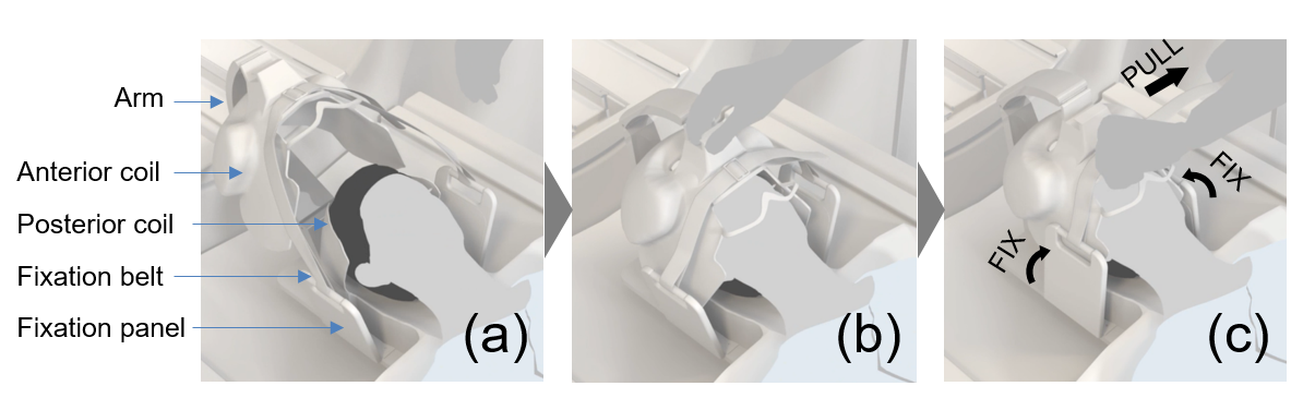
Setting of a size-adaptable tight-fit head array.
(a) Firstly, the patient lies in the posterior coil. Here, the flexible
anterior coil is supported with an arm where it does not interfere with the
patient. (b) Secondly, the technician slides the arm to set the anterior coil to
the patient. (c) Lastly, the technician pulls the fixation belt for a
one-action fixation. Here, the flexible anterior coil tightly fits to the
patient’s head, and the fixation panels closes in for a firm head fixation.
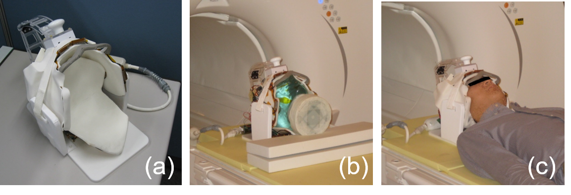
Prototyping of a size-adaptable tight-fit
head array. (a) 16-channel size-adaptable coil prototype. (b) Size-adaptable
coil with a head-shaped phantom. (c) Size-adaptable coil with a healthy
volunteer.
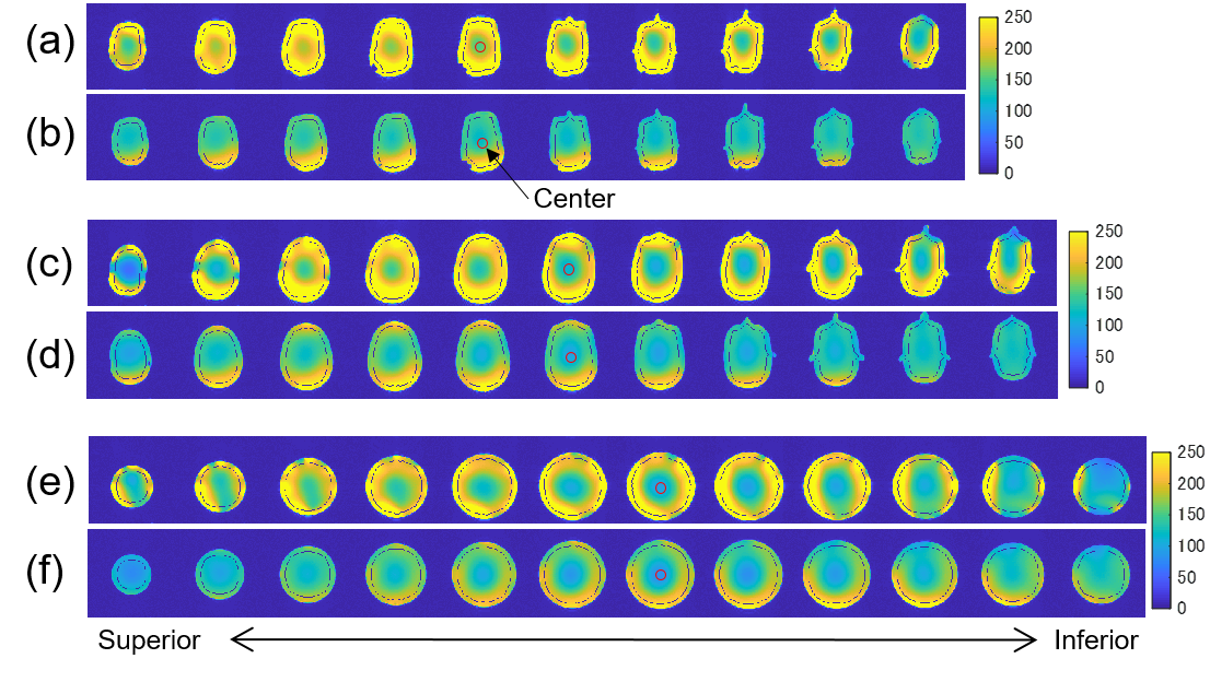
SNR maps of (a, c, e) a 16ch size-adaptable
coil, and (b, d, f) a conventional 15ch rigid coil, for phantoms of three
sizes, (a, b) small (phantom S), (c, d) large (phantom L), and (e, f)
extra-large (phantom XL). Perimeters of each phantoms were 514 mm, 591 mm, and
619 mm, respectively, which covers the range of 1 to 99 percentile of adult
head circumference. Two regions of interest are shown; (1) Whole brain region:
8 voxels shrunk from the edge, (2) Center region: 30 mm diameter at the center
of the middle slice (red circle).
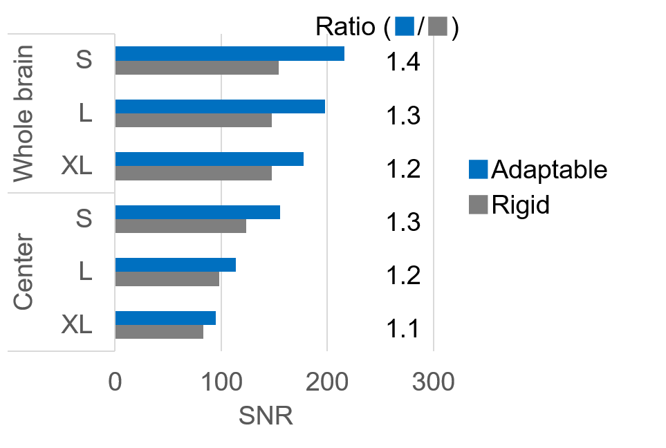
Average SNR of two regions of interest
(whole brain region and center region) for phantoms S, L, and XL. Ratio of the
average SNR or the size-adaptable coil against the rigid coil is shown on the
right of the bar charts.
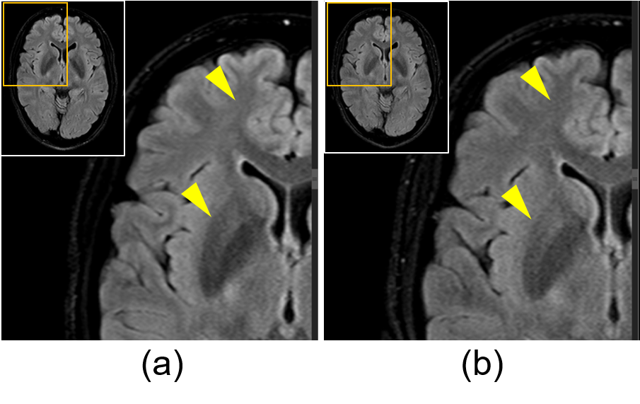
FLAIR
image of a healthy volunteer with (a) a 16ch size-adaptable coil, and (b) a
conventional 15ch rigid coil. The enlarged region is shown with an orange box.