4039
Development of variable-temperature MR microimaging system with a large-diameter solenoid coil for a vertical wide bore superconducting magnet1Institute of Applied Physics, University of Tsukuba, Tsukuba, Japan, 2Department of Forest Science, Graduate School of Agricultural and Life Sciences, The University of Tokyo, Tokyo, Japan
Synopsis
We have developed a large-diameter, solenoid probe for a wide bore NMR superconducting magnet that can image samples at different temperatures. The controllable temperature range was -20 to 20 °C. To demonstrate the performance, plant buds were imaged at different temperatures, and the frozen process was visualized.
INTRODUCTION
NMR microimaging at variable temperatures has many applications, such as plant imaging and cryobiopsies. A conventional variable-temperature probe uses a saddle RF coil that has a lower SNR than a solenoid coil, or uses a solenoid RF coil but with a limited sample area (<10 mm). In this study, we developed a variable-temperature microimaging system with a 40 mm-diameter solenoid coil for large sample observations. This system used a planar gradient rather than a saddle one in order to provide a large space for samples. The temperature was controlled by air and the whole system was simple, which reduced the running and maintenance costs. We demonstrated microimaging of plant samples during the freezing process, and showed the performance of the system.METHOD
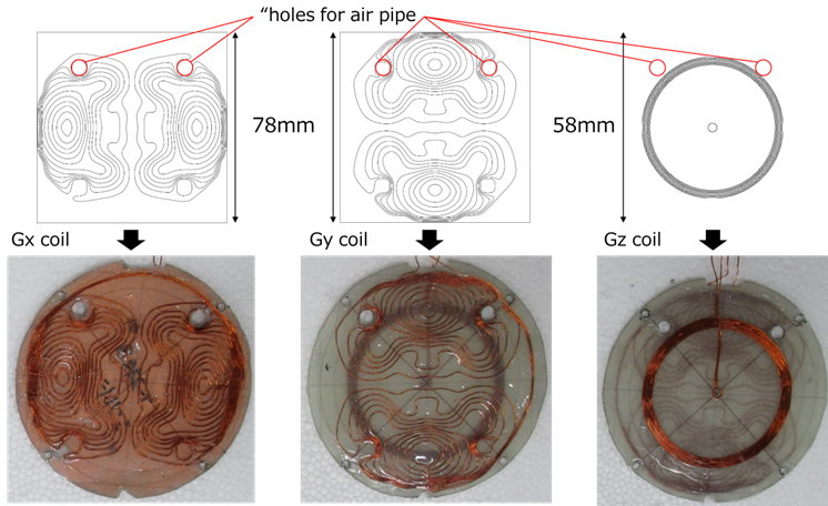
The variable temperature RF probe was designed for a 4.74 T/89 mm horizontal bore superconducting magnet (Oxford Instruments plc, abingdon, UK). It consisted of a planar gradient coil set, solenoid RF coil, and acrylic sample holder (the inner space = 38 mm, length = 65 mm) (Figure 1). The solenoid RF coil (inner diameter = 40 mm , length = 40 mm, 4 turns) was wound using a 1.5 mm polyurethane-coated Cu wire, and divided into 4 segments by using 3 chip capacitors (4.8 pF) to achieve 50 Ω impedance matching at 202 MHz.The planar gradient coil (Fig. 2) were designed and fabricated as follows. The coil planes had holes (7 mm in diameter) through which air pipes for cooling passed. The x and y gradients were designed using GA-DUCAS 1 (homogenous area = 40 mm diameter of spherical volume (DSV), coil plane diameter = 78 mm). The z gradient designed using an artificial bee colony algorithm 2 (homogenous area = 40 mm DSV, coil plane diameter = 58 mm). The gradient coil was wound on FRP plates using polyurethane coated Cu wire (wire diameter = 0.4 mm). The efficiencies were 0.60 (x), 0.51 (y), 0.72 (z) G/cm/A, respectively.
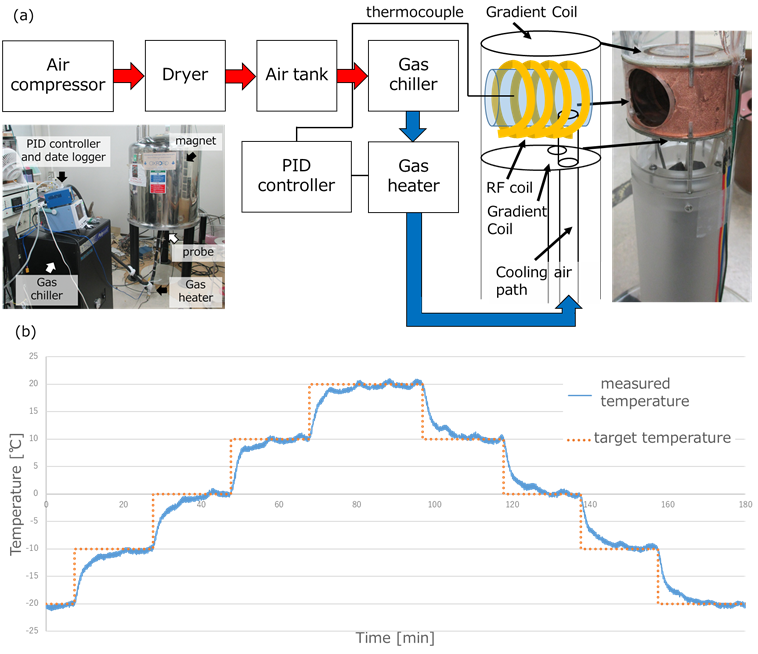
The base metal of the probe was made of acrylic instead of metal to avoid the eddy current field caused by gradient switching. Figure 3(a) shows a diagram of the cooling system. Dried air from a dryer was stored in a tank, and cooled by a gas chiller. Then the temperature of the flowing air was kept constant using a PID controller and gas heater. All experiments were performed on a home-built digital MRI console (DTRX6, MRTechnology, Tsukuba, Japan). Flower buds, Aesculus turbinate and Rhododendron, were imaged at different temperatures using a 3D spin echo sequence (TR / TE = 800ms / 9ms, FOV = 51.2 mm × 25.6 mm × 25.6 mm, image matrix = 512 × 256 × 64, voxel size = 100 um × 100 um × 400 um).
RESULTS AND DISCUSSION
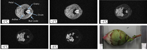
Figure 3(b) shows a temperature change in the empty sample holder. The temperature was controllable ranging from -20 ℃ to 20 ℃. The temperature fluctuation after reaching the steady state was within 0.8 °C at the target temperature of -10 ℃. Figure 4 shows MR images of the flower bud acquired at 1, -3, -7, and -11 ℃. The signals of florets and bud scales decreased as the temperature decreased. This indicates that these tissues were frozen gradually within this temperature range. The xylem was not frozen at -11 ℃ and was clearly visible in the image. This agrees with the fact that xylem tissues are supercooled and hardly frozen in many plants 3. Figure 5 shows MR images of the flower bud, Rhododendron, acquired at 0, -2, -4, -6, and -8 ℃. The bud scales and the other tissues were frozen below -4℃, while the anthers were partly frozen. The ovules were hardly frozen, which is the first observation for Rhododendron.CONCLUSION
We developed a variable temperature MR microimaging system using the large-diameter solenoid coil for the vertical wide bore superconducting magnet. We imaged plant flower buds at the low temperatures and demonstrated the system performance.Acknowledgements
No acknowledgement found.References
1. K. Matsuzawa et al., A new method for optimizing performances of gradient coils based on singular value decomposition and genetic algorithm, 2017, 25th Annual Meeting & Exhibition (ISMRM),Honolulu, USA.(4336)
2. Y. Terada et al., A Straightforward Direct Optimization Method for Designing Biplanar Gradient Coils Using Artificial Bee 3olony Algorithm, 2015, 23th Annual Meeting & Exhibition (ISMRM), Toronto, Canada.(1834)
3. M. Ishikawa et al., Freezing behaviours in wintering Cornus florida flower bud tissues revisited using MRI, plant, Cell and Environment. 2016;2663-2675.
Figures