4033
RF endoluminal coils with NMR electro-optical conversion and transmission for colon wall imaging: Initial finding
Paul Nobre1, Alice Ferrando1, Gwenaël Gaborit2,3, Raphaël Sablong1, and Olivier Beuf1
11Univ. Lyon, CREATIS ; CNRS UMR 5220 ; INSERM U1206 ; INSA-Lyon ; UJM-Saint Etienne ; Université Lyon1 ; 69616 Villeurbanne, France, Lyon, France, 2University of Savoie, IMEP-LAHC, UMR 5130, 73376 Le Bourget-du-Lac, France, Le Bourget-du-Lac, France, 3KAPTEOS, 73376 Sainte-Hélène-du-Lac, France, Sainte-Hélène-du-Lac, France
11Univ. Lyon, CREATIS ; CNRS UMR 5220 ; INSERM U1206 ; INSA-Lyon ; UJM-Saint Etienne ; Université Lyon1 ; 69616 Villeurbanne, France, Lyon, France, 2University of Savoie, IMEP-LAHC, UMR 5130, 73376 Le Bourget-du-Lac, France, Le Bourget-du-Lac, France, 3KAPTEOS, 73376 Sainte-Hélène-du-Lac, France, Sainte-Hélène-du-Lac, France
Synopsis
Receive surface coils in MRI have proven to enable higher spatial or temporal resolution. However, the proximity of the galvanic connexions with the body when the coil is used as receiver coil together with a volume coil can lead to safety issues, in particular for inner coil. This configuration can induce currents in coaxial cable and local SAR increase can be induced. Replacing the galvanic connexions with optical ones could prevent those risks and enable safe colon wall imaging with endoluminal coils. First experimental demonstration of an electro-optic conversion based on Pockel’s effect is demonstrated at 4.7T.
Introduction
Higher spatial resolution MRI of the colon wall could lead to diagnosis and characterization of colon cancer at an earlier stage thus increasing the five years survival rate. Endoluminal coils increase the image spatial resolution at close proximity of the loop coil, therefore allowing a better analysis of the colon wall1. Nonetheless, the coaxial cable connecting the coil to the MR system causes safety issues2, as the excitation radiofrequency magnetic field B1 induces currents in the galvanic connexion leading to a local increase of the SAR. This unwanted effect can be avoided by using optical fiber instead of metallic cable3. For that purpose, the NMR signal must be converted into light signals and converted back in electrical signal to be digitalized and reconstructed by the MR system. This work presents the first MR images obtained on a preclinical MR system after electro optical conversion using Pockel’s effect.Methods
Endoluminal coils previously designed and built for mouse colon imaging were used1. The coil loop was 40mm in length and 1.6mm in width and was operating at 200.29 MHz corresponding to the proton frequency of the 4.7T preclinical MRI scanner used (Bruker, Germany). The endoluminal receiver RF output signal of the coil was connected to a MAXN-LN-20 (IXblue, France) used as electro-optical converter. The variations of the electrical field change the polarization of the 1550nm linearly polarized laser going through the crystal by Pockel’s effect. An eoSense system (Kapteos, France) was used as the opto-electronic converter. To asses SNR, a tube was filled with saline solution doped with nickel sulphate (1.25 g/L of NiSO4 + 5 g/L of NaCl) in order to reduce T1-value. The coil was inserted inside the tube and sealed. A 72mm inner diameter birdcage coil (Rapid Biomedical, Rimpar, Germany) was used as transmit coil. The figure 1 represents the optical transmission chain. MSME sequence with TR/TE 650/14ms was performed with optical conversion and compared with with galvanic connexions and a quadrature 32mm inner diameter birdcage coil dedicated to mouse.Results
As predicted, the surface coil with galvanic connexion depicts a considerable gain in SNR compared to the volume coil. As seen in the figure 2, SNR profiles obtained with the galvanic transmission chain is 20 times better that the optic one. However, there is a clear correlation between both SNR profiles. Despite the fact that achieved EO conversion do not reach the targeted SNR, the SNR in the area of interest (close proximity of the loop) is still higher than with the dedicated volume coil (figure 3).Discussion
As predicted, the SNR obtained with the galvanic connexion endoluminal coil has an excellent SNR close to the loop coil (target colon area), making it a good objective reference. The proof of concept of the conversion chain based on Pockel’s effect was demonstrated. Beside the fact the image is noisier, the figure 4c depict a similar image in shape than figure 4b. The SNR of the optical coil is however significantly inferior to the galvanic one but above the SNR obtained with the volume at the wall of the phantom. One lead could be to improve the linearity of the converter. It depends on the operating point defined by a voltage offset, which was not studied in depth for this test. Secondly, the bandwidth of the electro-optical converter was not optimal for this application and the geometry of the crystal should be the subject of further improvements. Developing application-specific converter with suited bandwidth and operating point could improve significantly the SNR.Conclusion
The concept of EO conversion based on a bulk crystal using Pockel’s effect was demonstrated with an endoluminal coil for colon mouse. The next step is the improvements of the SNR and to add an efficient optical-based decoupling circuit as already demonstated3. This, in addition to monitoring the induced currents with an optoelectronic Electrical field probe4 would pave the way to in vivo testing.Acknowledgements
This work funded by the AURA region is performed within the scope of LABEX PRIMES (ANR-11-LABX-0063) of Université de Lyon, within the program "Investissements d'Avenir" (ANR-11-IDEX-0007) operated by the French National Research Agency (ANR). Experiments were performed on the PILoT facility, part of the France Life Imaging infrastructure (ANR-11-INBS-0006).References
1. H. Dorez et al. (2016), Endoluminal high-resolution MR imaging protocol for colon walls analysis in a mouse model of colitis, Magn. Reson. Mater. Phys. Biol. Med., vol. 29, no 4, p. 657-669. 2. V. Detti, et al. Magnetic Resonance in Medicine, vol. 66, p. 448-455, 2011. 3. I. Saniour et al. (2017). Active optical-based detuning circuit for receiver endoluminal coil. Biomedical Physics & Engineering Express, vol 3 no 2. 4. I. Saniour et al. (2017). Electro-optic probe for real time assessments of RF electric field produced in an MRI scanner: Feasibility tests at 3 and 4.7 Tesla. NMR in Biomedicine. Vol 31. No 1.Figures
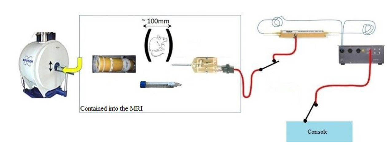
Fig. 1. Diagram of optical
conversion chain. Inside the MRI from left to right is the emission coil, the
phantom simulating the mouse’s colon and the endoluminal coil. Outside are the
converters and the console processing the images.
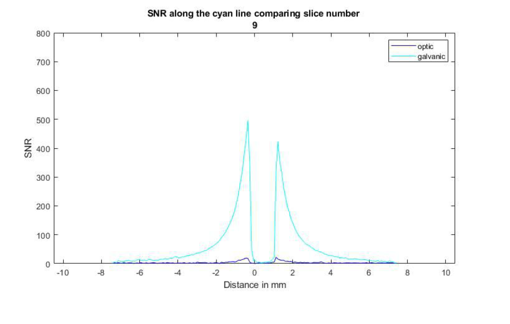
Fig. 2. Comparison of the local SNR for the endoluminal coil with galvanic cable
(in cyan) and with optical fiber. The slice number 9 was chosen because it was
in the middle of the phantom where the distribution of the liquid was uniform.
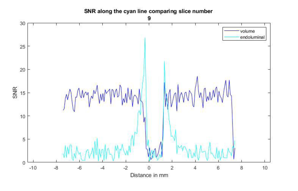
Fig. 3. Comparison of the local SNR between volume coil reception (in dark blue) and
endoluminal coil with optical fiber. While the volume coil has higher average
SNR, the endoluminal is almost 2 times better at the tube’s walls.
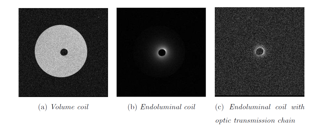
Fig. 4. Images of the saline solution tube obtained with the different reception
coils. For reference, the inside of the tube is 1.6mm