4029
An Implantable MRI Micro-coil of Blood vessel for High Sensitivity and High SNR Imaging at high field MRI.1Department of Biomedical Engineering, Hanyang University, Seoul, Republic of Korea
Synopsis
The importance of implantable micro-coil has been rapidly increased due to its advantages in diagnosing intravascular and brain diseases. In this study, the receive-only conformal-shaped micro-coil was designed and presented. Our research demonstrated that homogeneous B1- magnetic field distributions and receptive sensitivity around the blood-vessel walls were achieved by using the proposed micro-coil.
Introducation
Surface coils are in close vicinity of the human body to detect the high sensitivity image of the target tissues, but some deep blood vessel tissues are not detected efficiently by the surface coil [1]. The alternative method to visualize the blood vessel walls is to use the implantable receive-only micro-coil [2-4]. Most of the previous studies used single loop coils, solenoid coils, tilted coils and meandering line for receiving coils. However, most of the designs had limited success and suffered from a number of problems. Usually, the coil's most sensitive area is located in the arterial not the artery wall, and severe image artifacts are often found during intense blood flow due to the coil's movement. This paper proposed a new conformal-shaped micro-coil inside the blood vessel to improve the blood-vessel wall imaging and sensitivity.Methods
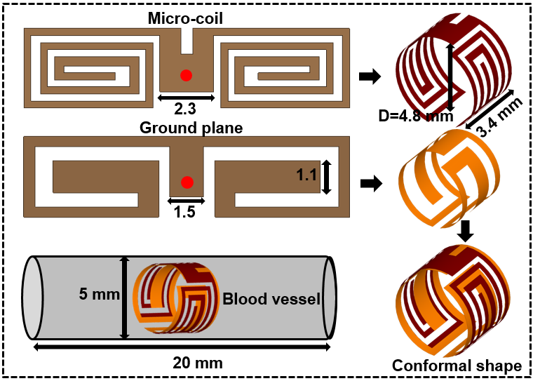
This study designed a small size receive-only micro-coil for blood vessel imaging. The coil resonates at 7 T (300 MHz) and 3 T (128 MHz) by inserting and controlling the trimmer capacitor. To analyze the performance of the proposed micro-coil, B1- field distributions around the blood vessels were calculated by finite-difference time-domain (FDTD) method using the Sim4Life simulator by ZMT [5]. As shown in Fig. 1, the conformal shape micro-coil is placed in the vicinity of blood vessels and muscle-mimicking phantom. The proposed coil was compared with the surface coil, solenoid coil, and body Birdcage coil to validate the field sensitivity of the coil for various coil arrangements. To examine the performance of micro-coil in a realistic environment, the proposed coil was implanted in the blood vessel of brain, neck, kidney, and heart of "Duke" model in Sim4Life, which provides an efficient modeling of detailed anatomical parts of the human body. For simulation results verification, the proposed coil was fabricated and placed in the blood vessel and ASTM models to measure the S-parameters.Results
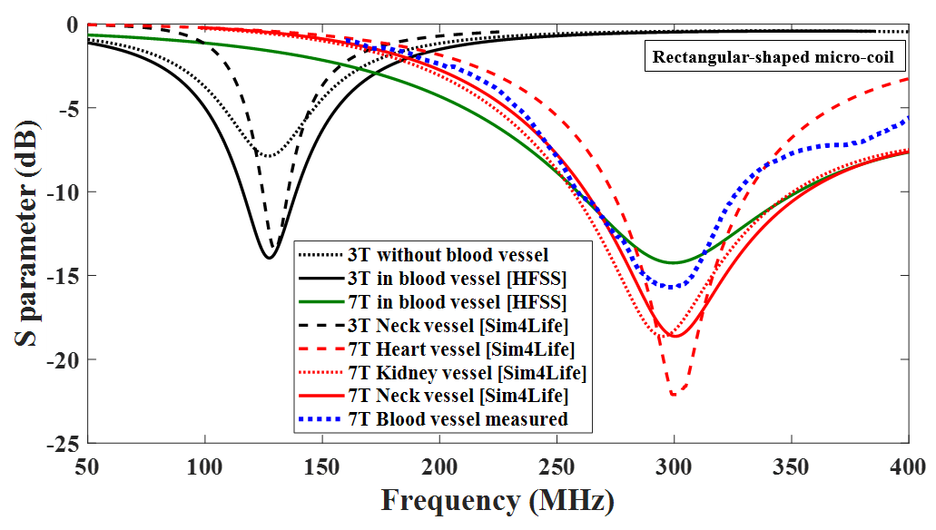
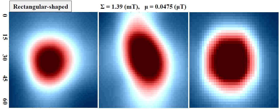
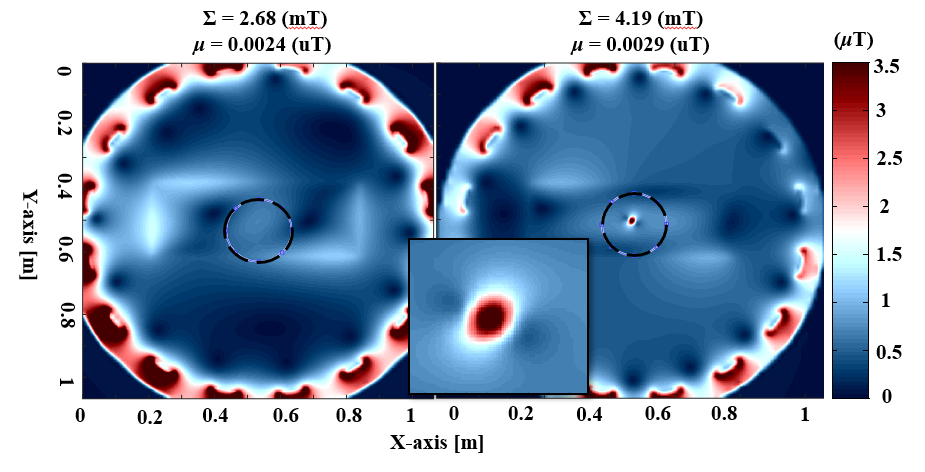
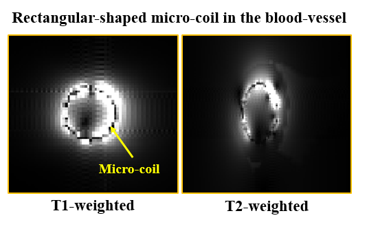
The open interior of the coil provides the advantage of no blood flow abstraction and the conformal shape gives symmetric field distribution, which is helpful for high sensitivity imaging around the blood-vessel walls. Fig. 2 illustrates the simulated scattering parameters (S11) of the micro-coil within the dielectric phantom and human duke model at both 3 T and 7 T, and the coil is fully tuned and matched at 7 T and 3T MRI. Fig. 3 shows that the field distributions are improved and the uniform B1- fields are pronounced in the blood vessel. In the region of interest (ROI), the reception efficiency and the sum of the B1- fields improved 75% and 72 %, respectively. It can be observed from the B1- distributions of the coil that the high central magnetic fields are clearly pronounced in the center of every slice, while they become weaker as the distance increase away from the micro-coil. Fig. 4 shows the comparison of simulated B1- fields in the central axial slices within the ASTM phantom for the combined approaches between the Birdcage coil and the micro-coil. The B1- field distributions using a combined approach were pronounced higher in the ROI compared to the Birdcage coil only. Fig. 5 shows the MR images of central axial slice for the micro-coil at blood vessels. It can be seen from the figure that the micro-coil allowing the selective imaging of the arterial walls and the resulting sensitivity is relatively homogenous around the coil inside the blood vessel.Conclusion
The proposed coil can be used for smaller deep blood vessel MRI imaging. The small size of the micro-coil makes the placement of the coil possible inside the blood vessel. This study suggests that the micro-coil in the immediate vicinity of the plaque with the coil receptive area limited to the ROI only will deliver the best possible factor for plaque and MR intracranial aneurysm scanning. The proposed coil operates efficiently in different blood vessel environments.Acknowledgements
This work was supported by the Basic Science Research Program through the National Research Foundation of Korea (NRF) funded by the Ministry of Science and ICT under Grant 2019R1A2C2004774.
Sim4Life by ZMT, www.zurichmedtech.com
References
1. Atalar E, Bottomley PA, Ocali O, Correia LC, Kelemen MD, Lima JA, Zerhouni EA. High resolution MRI and MRS by using a catheter receiver coil. Magnetic resonance in medicine. 1996 Oct364
2. Farrar CT, Wedeen VJ, Ackerman JL. Cylindrical coil for magnetic resonance studies of atherosclerotic plaque. Magnetic Resonance in Medicine. 2005 Jan531
3. Visser HJ, den Kamp NA, Aben MJ, Seppenwoolde JH, Bartels LW, Bakker CJ, Tijhuis AG. An analytical model for MR antennas. (2007): 589-589
4. Anderson KJ, Leung G, Dick AJ, Wright GA. Forward‐looking orthogonal‐solenoid coil for imaging and guidance in occlusive arterial disease. Magnetic Resonance in Medicine. 2008 Aug602
5. Sim4Life by ZMT, https://www.zmt.swiss.
Figures