4019
A Novel Clinical Friendly 7T T/R 32-Channel Head Coil Using Skipped-Rung Birdcage as Transmitter
Tsinghua Zheng1, Xiaoyu Yang1, Blaise Whitesell1, Martin Domondon1, Paul Taylor1, Samuel Musilli1, and Labros Petropoulos1
1Quality Electrodynamics, LLC, Mayfield Village, OH, United States
1Quality Electrodynamics, LLC, Mayfield Village, OH, United States
Synopsis
We propose a novel skipped-rung birdcage coil as the local transmitter for head imaging at 7 Tesla. This concept allows sizable openings on the coil anterior. The openings improve both patient comfort and workflow. A 7T head coil using the skipped-rung birdcage transmitter and a 32-channel receiver array was constructed and evaluated. The preliminary test results show a good transmitting efficiency and a good image quality compared with a commercial 7T head coil.
Introduction
Birdcage coil has been used as a volume transmitter for its uniform B1 profile and good transmit efficiency at 3T and below(1). The birdcage coil transmitting efficiency is affected by the coil loss, the sample loss and the radiation loss. At ultra-high frequency (UHF), such as 7T and above, its transmitting efficiency reduces significantly due to the excessive radiation loss. This is because the radiation power Pave is proportional to the square of the frequency f(2). PAve=4πf2μεE0(r)H0(r) The amplitude of the RF current through each rung of a birdcage may be written as Irung= I0/N, where I0 is a scaling factor from the targeted B1 field strength. The total radiation power P may be the sum of all rungs that can be written as P~(I0/N)2*N=I02/N. Thus, one approach for reducing radiation loss of the 7T birdcage coil is to increase the number of rungs. In many previous 7T head coil designs, the number of rungs for the birdcage transmitter is equal or greater than16(3)(4)(5). In addition, an RF shield must be placed closely outside the birdcage for further reduction of radiation loss. The obvious drawback of more rungs and the RF shield is that it is becoming impossible to have openings large enough for eyes on the anterior of the coil. The eye openings are crucial for reducing claustrophobic reactions and facilitating visual stimulation for 7T head applications. Therefore, the head imaging experiences at 7T in terms of patient comfort and workflow are suffering. We address this issue by proposing a novel birdcage coil. The novelty is to strategically remove a small number of rungs in the birdcage coil and the shield near eyes, and create openings on the mechanical housing. The B1 field will have local disturbance at where the rungs are removed. However, the overall B1 uniformity across the whole imaging volume may not be affected significantly. We call this concept a skipped-rung birdcage.Method
B1 uniformity evaluation was performed using EM simulation software Remcom XFtdt. A birdcage coil with a partial shield model was created with the following parameters: diameter=30cm, length=22cm, partial shield diameter=33cm, frequency=297 Mhz. Two configurations were evaluated: (1) conventional birdcage coil with evenly distributed 24 rungs, and (2) the same dimension birdcage coil except two rungs adjacent to the top center rung are removed. The ring currents for both configurations are the same. Fig.1 shows the simulation results of B1 profiles for both configurations using a 20cm radius no-load phantom. The result shows the B1 uniformity within the phantom for both cases are almost identical. We conclude that the removal of 2 rungs on the 24-rung birdcage has minimal impact on uniformity. Then a 7 Tesla T/R head coil with a skipped-rung birdcage as the transmitter and a 32-channel receive array was constructed. Fig. 2 shows the section of the birdcage coil where the rungs) were removed to create openings. A partial shield(6) was implemented for further reduction of radiation loss. The receiver array was constructed on the same mechanical housing under the transmit coil. It consists of 8 rows of 4 overlapped receive-only coils. 4 rows are in the anterior and 4 are in the posterior. Each coil is connected to a low impedance preamplifier to reduce coupling with its neighboring coils. Fig. 3 shows the anterior and posterior housing of the 7T T/R 32 channel head coil. Besides the two eye openings at the top, there is another opening at the superior end of the coil that allows the EEG probes to go through. In addition, the internal profile of the housing has mechanical cutouts to accommodate headphones. Comparing to a commercial 7T head coil (Nova Medical), the split-top design makes the workflow much easier. Even with larger inner diameter, the new coil overall dimension is smaller (37cmx48cmx38cm) than the comparison 7T head coil (68cmx 48.5cmx 37.5cm). The coil is also 40% lighter (8kg vs.14kg).Results
The 7T T/R head coil with skipped rung birdcage was tested on a Siemens MAGNETOM 7T scanner. Axial GRE images show uniform B1 profile on a no-load spherical phantom, as shown in Fig. 4. The transmit adjustment voltage was 20% higher (250V vs. 210V) than the comparison coil. We attribute the higher transmit voltage to a longer S-I dimension of the birdcage, which extends the coverage to the upper c spine. Volunteer studies were conducted using the 7T T/R skipped-rung 32 channel head coil and the comparison 7T 32 head coil. Results show that the two coils have comparable image qualities with commonly used head imaging protocols, such as GRE, MP2RAGE and EPI. Fig. 5 shows an example of an MP2RAGE result for comparison.Conclusions
This novel coil concept allows the eye openings on the anterior of the coil. The openings make the coil more patient friendly for head imaging at 7T. Preliminary volunteer evaluation has demonstrated a good transmitting efficiency and a comparable image quality as a commercial 7T 32 channel head coil. To further optimize the skipped-rung birdcage concept, our future work may explore various configurations of the rungs for the balance among the coil opening, the transmitting efficiency, and the B1 uniformity.Acknowledgements
The authors are grateful to Dr. Mark Lowe, Dr. Ken Sakaie and Dr. Sanghoon Kim at Cleveland Clinic Foundation (CCF) and Dr. Ravi Seethamrazu at Brigham Women’s Hospital (BWH) for their Siemens 7T MR system supports.References
(1) C.E. Hayes et al, An efficient, highly homogeneous radiofrequency coil for whole-body NMR imaging at 1.5 T, J. of Mag. Reson. Vol 63, Issue 3, 622-628 1985 (2) M. Muhibbullah et al, Frequency dependent power and energy flux density equations of the electromagnetic wave, Results in Physics Vol. 7, Pages 435-439, 2017 (3) P.J. Ledden, et al., 32 Channel Receive-Only SENSE Array for Brain Imaging at 7T, Proc. Intl. Soc. Mag. Reson. Med. 15 2007. (4) G.C. Wiggins, et al., A 32 Channel Receive-only Head Coil and Detunable Transmit Birdcage Coil for 7 Tesla Brain Imaging, Proc. Intl. Soc. Mag. Reson. Med. 14, 415 2006 (5) M. Finnerty et al, A 7-Tesla Transmit with 32-Channel Receive-Only Array Head Coil for fMRI, Proc. Intl. Soc. Mag. Reson. Med. 2016-2133 2016 (6) M. Finnerty et al, A 7-Tesla High Density Transmit with 28-Channel Receive-Only Array Knee Coil, Proc. Intl. Soc. Mag. Reson. Med. 2010-642 2010Figures
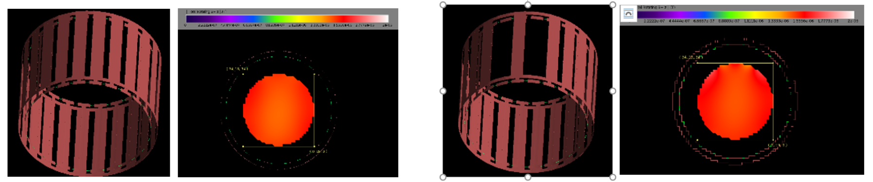
Fig.1 EM simulation model and B1 profile of a 24-rungs birdcage coil
(left) and a skipped-rung (22 rungs) birdcage coil (right)
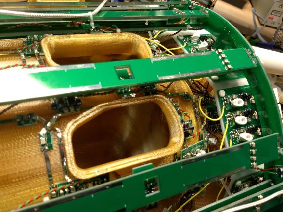
Fig. 2 Structure of the skipped-rung birdcage and the eye openings
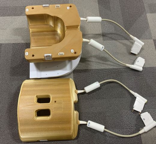
Fig. 3 Anterior and posterior parts of the 7T T/R skipped-rung 32-channel
head coil
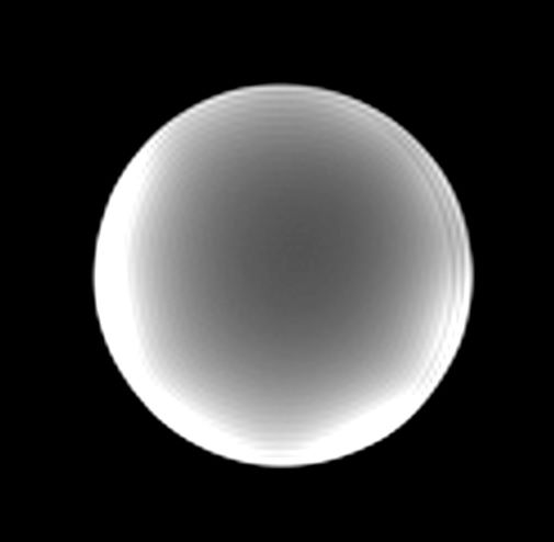
Fig. 4 Axial GRE images of 7T T/R skipped-rung 32-channel head coil using
a spherical no-load phantom
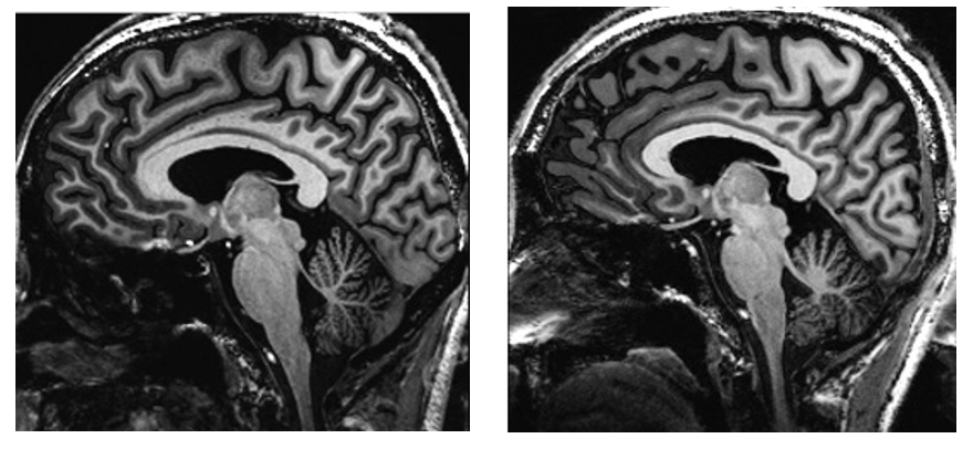
Fig. 5 MP2RAGE images of the 7T T/R skipped-rung 32-channel head coil
(left) and the commercial 7T 32 channel head coil (right)