4018
A tight-fit flexible high-impedance coil array for high-resolution imaging of small ex-vivo specimen using a human 7T scanner1Human Brain Research Center, Graduate School of Medicine, Kyoto University, Kyoto, Japan, 2Center for Advanced Imaging Innovation and Research (CAI2R) and Bernard and Irene Schwartz Center for Biomedical Imaging, Department of Radiology, New York University School of Medicine, New York, NY, United States, 3Department of Neurology, Kyoto University Graduate School of Medicine, Kyoto University, Kyoto, Japan, 4Department of Human Health Sciences, Kyoto University Graduate School of Medicine, Kyoto University, Kyoto, Japan
Synopsis
By borrowing concepts from the high-impedance glove coil, we propose an “elastic-tube coil” for imaging ex-vivo specimen at 7 Tesla. The elasticity of the sock makes it easy to setup and ensures a minimal distance between the coils and the specimen. Two high-impedance elements were stitched to an elastic tube suspended in a custom 3D printed housing. Volumetric gradient echo images of an ex-vivo human brain specimen were acquired with 60um-isotropic resolution.
Introduction
Quantitative comparison has shown that an approximately 3x higher SNR can be achieved at 7T compared to 3T (1). To reap these benefits, appropriate coils must be used. There are a number of researches in the high-resolution scan for ex-vivo small specimen of human brain (2-4). Ex-vivo whole-brain scan with 7T is also a target of research (5). When imaging ex-vivo samples of different sizes and shapes one would either need multiple coils or a scalable solution (6). High-Impedance coil (HIC) elements can provide great flexibility and robust decoupling (7). When using off the shelf 50-ohm coaxial-cable, the size of a HIC element shrinks to ~3.5cm in diameter. Considering that the depth at which a coil element is most efficient is comparable to its diameter (8), 50-ohm coaxial-cable based HIC elements are most suitable for samples up to ~7cm in size, such as ex-vivo specimen. However, based on our experience from a previous design, we found that the relatively large peripheral circuits (such as matching board and cable traps) can be fragile and time-consuming to set up. In this work, we endeavor to overcome these challenges by mounting the elements on an elastic tube, thus creating an easy to use setup while ensuring the minimal distance between the coil distance and specimen.Methods: coil design
Our ex-vivo coil consists of home-built birdcage coil for transmit and a two-channel HIC array for receive. The birdcage coil was constructed from copper tape placed on a 150mm diameter acrylic pipe. The coil consists of 8 rungs with a length of 150mm with a PIN diode on each of them for detuning. The coil was driven in a quadrature mode through a quadrature hybrid coupler. Reception, for use without the HIC insert, is also possible through the use of T/R switches. Our HICs were built from micro-coax (∅=1.1mm). The schematic design of the HIC and its physical implementation is shown in Figure 1. Two PIN diodes were used to detune the HIC during transmit. For each coil, a small copper shield was placed around the matching inductor to avoid coupling with adjacent coils. The cable length between the matching circuit and the preamplifier was adjusted to achieve “reversed” preamplifier decoupling (7). For each coil element, one cable trap was inserted between the coil and the preamplifier.Methods: tube holder design
To protect the HIC array while preserving its size adjustability, a dedicated tube holder was designed using Fusion 360 (Autodesk, USA) and was printed using a commercial Fused Deposition Modeling (FDM) 3D printer. The elastic tube holding the HIC elements was made from a sock by cutting off one end. A ring-like structure with a diameter of 53mm was designed to tightly hold one end of the sock in place. The tube holder was designed such that the sample tube stays stable while saving space for coil elements and their peripheral circuits. The complete assembly and its components are shown in Figure 2.Methods: ex-vivo sample
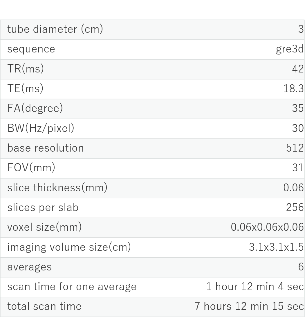
An ex-vivo brain specimen of an Alzheimer’s patient was scanned. The sample was immersed in a 20% sucrose. Prior to the scan at MRI, the sample was put into a tube with a diameter of 3cm with a 0.01M phosphate-buffered salts. (Figure 4).Methods: scan setting
All experiments were performed on an investigational whole-body 7T scanner (MAGNETOM 7T, Siemens Healthineers, Erlangen, Germany) equipped with body gradients. The brain specimen was scanned with a 3D gradient echo sequence with 60um-isotropic resolution in one average and six averages. Details of scan parameters are summarized in Table 1.Results and Discussion
Representative images of the specimen are shown in Figure 3-4. To obtain reasonable SNR at 60um-isotropic, 6 averages were needed (~7 hours). Optimization of scan parameters is still an ongoing process. Interestingly, no shimming was not required for this setup, most likely because of the small size of the sample tube and straight alignment of the tube along the B0. In its current configuration, the optimal field of view in the z-direction was approximately 4.0cm. This could be expected because of the 3.5cm diameter of the coil. Therefore, the sample had to be carefully centered in the z-direction. Therefore, increasing the number of elements (especially in the z-direction) could make it even easier to set up the experiment and would provide further improvements in SNR.Conclusion
By using a custom-built birdcage coil for transmit in combination with two HIC elements, 60-um isotropic imaging for an ex-vivo specimen in a 3cm diameter tube was possible in a reasonable time. The dedicated tube holder for protection of the HICs makes it easy to set up experiments quickly without fear of damaging the small HIC elements.Acknowledgements
We thank Dr. Hideto Kuribayashi (Siemens Healthcare KK) for useful discussion.References
1. Pohmann R, Speck O, Scheffler K: Signal-to-noise ratio and MR tissue parameters in human brain imaging at 3, 7, and 9.4 tesla using current receive coil arrays. Magn Reson Med 2016;75:801-809
2. Augustinack JC, van der Kouwe AJ, Blackwell ML, Salat DH, Wiggins CJ, Frosch MP, Wiggins GC, Potthast A, Wald LL, Fischl BR: Detection of entorhinal layer II using 7Tesla [corrected] magnetic resonance imaging. Ann Neurol 2005;57:489-494
3. van der Kolk AG, Zwanenburg JJ, Denswil NP, Vink A, Spliet WG, Daemen MJ, Visser F, Klomp DW, Luijten PR, Hendrikse J: Imaging the intracranial atherosclerotic vessel wall using 7T MRI: initial comparison with histopathology. AJNR Am J Neuroradiol 2015;36:694-701
4. Strotmann B, Kögler C, Bazin PL, Weiss M, Villringer A, Turner R: Mapping of the internal structure of human habenula with ex vivo MRI at 7T. Front Hum Neurosci 2013;7:878
5. Edlow BL, Mareyam A, Horn A, Polimeni JR, Witzel T, Tisdall MD, Augustinack JC, Stockmann JP, Diamond BR, Stevens A, Tirrell LS, Folkerth RD, Wald LL, Fischl B, van der Kouwe A: 7 Tesla MRI of the ex vivo human brain at 100 micron resolution. Sci Data 2019;6:244
6. Urayama S-I, Zhang B, Fujimoto K, Okada T, Cloos MA. A modular 7T high-impedance array for ex-vivo imaging. In Proc Int Soc Magn Reson Med 2019, 1451. Montreal
7. Zhang B, Sodickson DK, Cloos MA: A high-impedance detector-array glove for magnetic resonance imaging of the hand. Nat Biomed Eng 2018;2:570-577 8. Roemer PB, Edelstein WA, Hayes CE, Souza SP, Mueller OM: The NMR phased array. Magn Reson Med 1990;16:192-225
Figures