4001
Altered brain language network in idiopathic peripheral facial paralysis patients with dysarthria
Wenwen Gao1, Xiaowei Han1, Haimei Li2, Yijiang Zhu3, Lei Du1, Li zhi Xie4, and Guolin Ma1
1China-Japan Friendship Hospital, Beijing, China, 2Fu Xing Hospital, Capital Medical University, Beijing, China, 3Anhui Provincial Hospital, Hefei, China, 4GE healthcare, China, Beijing, China
1China-Japan Friendship Hospital, Beijing, China, 2Fu Xing Hospital, Capital Medical University, Beijing, China, 3Anhui Provincial Hospital, Hefei, China, 4GE healthcare, China, Beijing, China
Synopsis
Dysarthria is a common symptom of facial paralysis (FP). The aim of this study was to investigate the functional alterations of the brain language network in FP patients with dysarthria using resting-state fMRI. We found that the functional connectivity between bilateral language regions was significantly decreased in these patients compared with healthy controls. The decrease of functional connectivity in the language network was positively correlated with the severity of oral paralysis in patients. To sum up, these data suggest that dysarthria caused by facial nerve paralysis may lead to a decrease of neural activity in the brain language network.
Introduction
Dysarthria is one of the common symptoms of facial paralysis (FP)1, characterized by slurred or slow speech that can be difficult to understand. However, the exact neural mechanisms that regulate the language alterations in these types of patients remain unexplored. Early identification of functional changes associated with brain language network in FP patients with dysarthria can help to understand the pathogenesis of the disease and provide early diagnosis and timely treatment. The aim of this study was to investigate functional alterations in the brain language network of early idiopathic peripheral FP patients with dysarthria using resting-state fMRI.Methods
A total of 45 cases with FP (left 22, right 23) and 34 healthy controls (HC) were recruited into this study. The data of patients with left-side FP and matched controls (17 cases) were flipped from left to right, and the brain regions were defined as ipsilateral and contralateral regions. The FC of 16 ROIs in classical language centers and regions that may be involved in language function were calculated (Figure 1). After identifying the differences of FC between the two groups, the correlations between altered FC and TFGS score of oral muscle movement in FP group were analyzed.Results
The FC between bilateral language regions was significantly decreased in FP group compared with HC group (P < 0.05). Furthermore, the ipsilateral inferior frontal gyrus, superior temporal gyrus, and middle temporal gyrus had significantly decreased FC in multiple brain regions. In addition, we found significant alteration in FC in the thalamus and cerebellum of patients with FP, which indicated that these two regions may be relevant for dysarthria in FP (Figure 2). The correlation analysis indicated that the decrease of FC was positively correlated with the severity of oral paralysis.Conclusion
Idiopathic peripheral FP with dysarthria induces several FC alterations in the brain language network. The severity of oral paralysis is associated with these functional alterations. This study provides evidence that dysarthria caused by facial nerve paralysis may lead to a decrease of neural activity in the brain language network. Early identification of these changes furthers the understanding of the pathogenesis of the disease, thus providing opportunity for early diagnosis and treatment.Acknowledgements
This study was supported by the National Natural Science Foundation of China (NSFC) (Nos. 81971585 and 81571641), and The National Key Research and Development Program of China (Nos. 2016YFC1307001).References
1. Basić-Kes V, Dobrota VD, Cesarik M, et al. Peripheral facial weakness (Bell's palsy). Acta Clin Croat 2013; 52:195-202.Figures
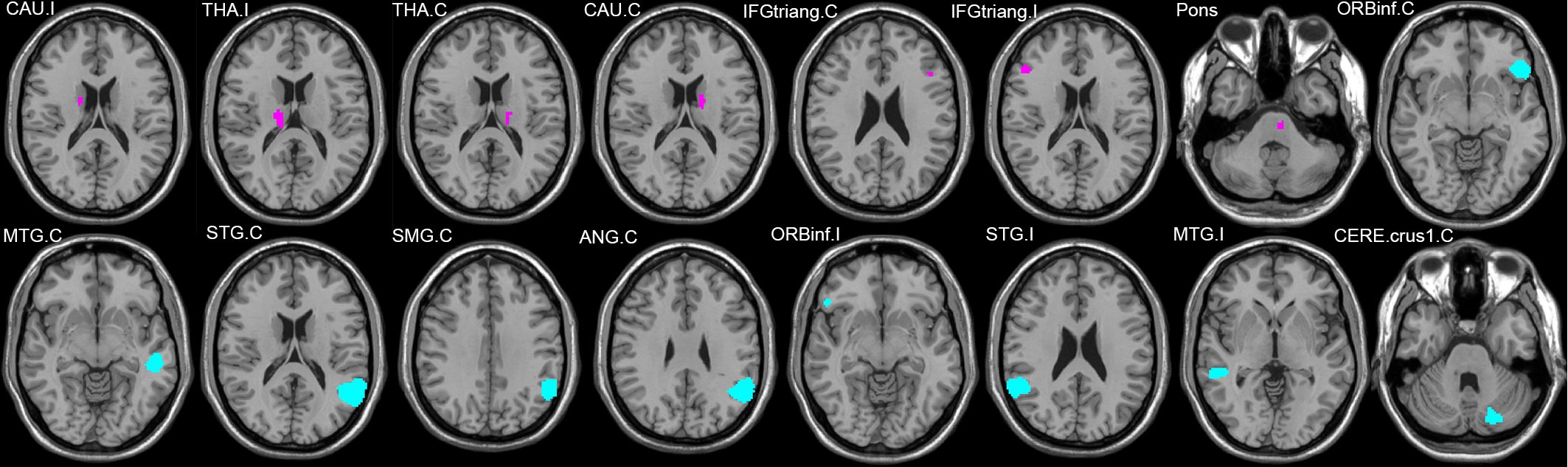
Fig.1
The ROI illustration of the basal
ganglia group (purple nodes) and the language group (green nodes) in the
Stanford University Willard 499 fROI atlas. The basal ganglia group and the language
group were selected because they cover the classical language centers and
regions that may be involved in language function (caudate, cerebellum,
thalamus, and pons).
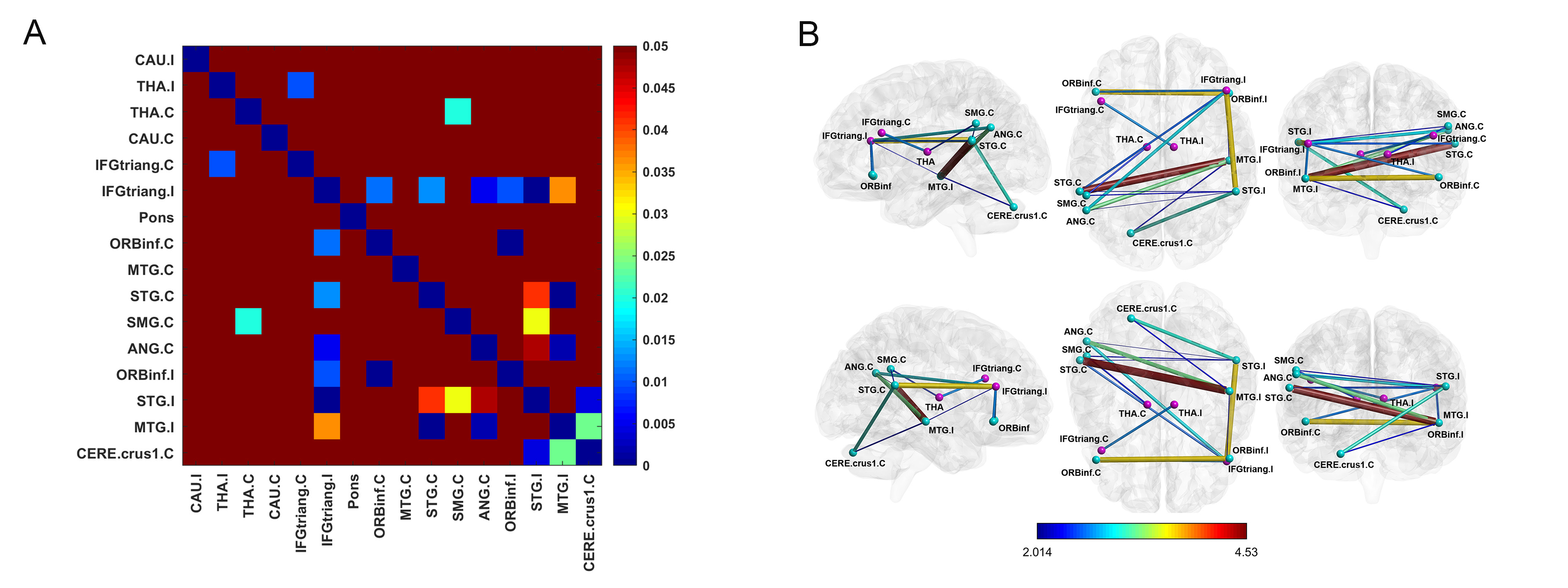
Fig.2
Two-sample T-test was conducted to
compare the differences in FC between the two groups; the threshold was set at
P < 0.05 FDR (false discovery rate). (A) The color grid
represents the P-value of each FC. (B) The brain regions and FC with
significant differences between groups; purple nodes represent the basal
ganglia group, green nodes represent the language group, while color and
thickness of edges represent the T values.