4000
Mapping chemotherapy-induced brain functional network abnormality in breast cancer survivors1Department of Medical Imaging and Radiological Sciences, Chang Gung University, Taoyuan, Taiwan, 2Department of Psychiatry, Chang Gung Memorial Hospital, Chiayi, Taiwan, 3School of Medicine, Chang Gung University, Taoyuan, Taiwan, 4Department of Diagnostic Radiology, Chang Gung Memorial Hospital, Chiayi, Taiwan, 5Medical Imaging Research Center, Institute for Radiological Research, Chang Gung University and Chang Gung Memorial Hospital at Linkou, Taoyuan, Taiwan
Synopsis
Chemo-brain is common among breast cancer survivors. However, some studies suggested cognitive function deficits may exist before chemotherapy initiation. Our study discovered the functional network alterations in women pre- (C-) and post-chemotherapy (C+) compared with health controls using resting-state functional MRI. The mfALFF showed changes in the prefrontal cortex, bilateral middle, right inferior temporal gyrus, right angular gyrus, left insula, and left caudate among three groups. Graph theoretical analysis demonstrated that the C+ group became inclined toward regular networks and the C- group became inclined toward random networks.
Introduction
Breast cancer is the leading cause of cancer-related death among women worldwide. Treatments for breast cancer including surgery, radiotherapy, adjuvant chemotherapy, targeted therapy and hormonal therapy. Due to the advance of medical technology, the mortality of breast cancer reduced dramatically, however, some patients with chemotherapeutic agent administration reported cognitive impairment after chemotherapy. This phenomenon is called chemo-brain or chemotherapy-induced cognitive impairment (CICI) which is common in breast cancer survivors. Some studies even suggested the deficits of cognitive function were prior to the initiation of chemotherapy 1. The aim of this study was to discover the functional network alterations in breast survivors pre- and post- chemotherapy using resting-state functional MRI (rs-fMRI).Methods
In this study, we recruited 172 female participants and separated into three different groups, including 57 breast cancer survivors whom finished chemotherapy between 3 to 12 months (C+), 45 breast cancer survivors without chemotherapy (C-) and 70 health participants (HC). Each participant was assessed on neuropsychological scales, including the Patient Health Questionnaire-9 (PHQ-9) for depression severity.In all 172 participants, fMRI data were size = 64 × 64, and voxel size = 3.4 × 3.4 × 4 mm3, and number of axial images = 31 were acquired using 3T MRI scanner (Verio, Siemens, Germany) at Chiayi Chang Gung Memorial Hospital. The rs-fMRI was performed in a gradient echo planner imaging sequence with the repetition time/echo time = 2000/30 ms, flip angle= 90°, number of excitations = 1, field of view = 220 × 220 mm2, matrix acquired to cover the entire brain volume. Each rs-fMRI run contained 300 image volumes, and the total scan time was approximately 10 min.
We obtained mean fractional amplitude of low frequency fluctuation (mfALFF) and graph theoretical topology from rs-fMRI to portray functional changes among three groups. Analysis of variance (ANOVA) and post-hoc two-sample t tests were implemented to compare mfALFF among groups using SPM12. Participants’ age and education level were considered as covariates and FDR-corrected p value of <0.05 was considered statistically significant.
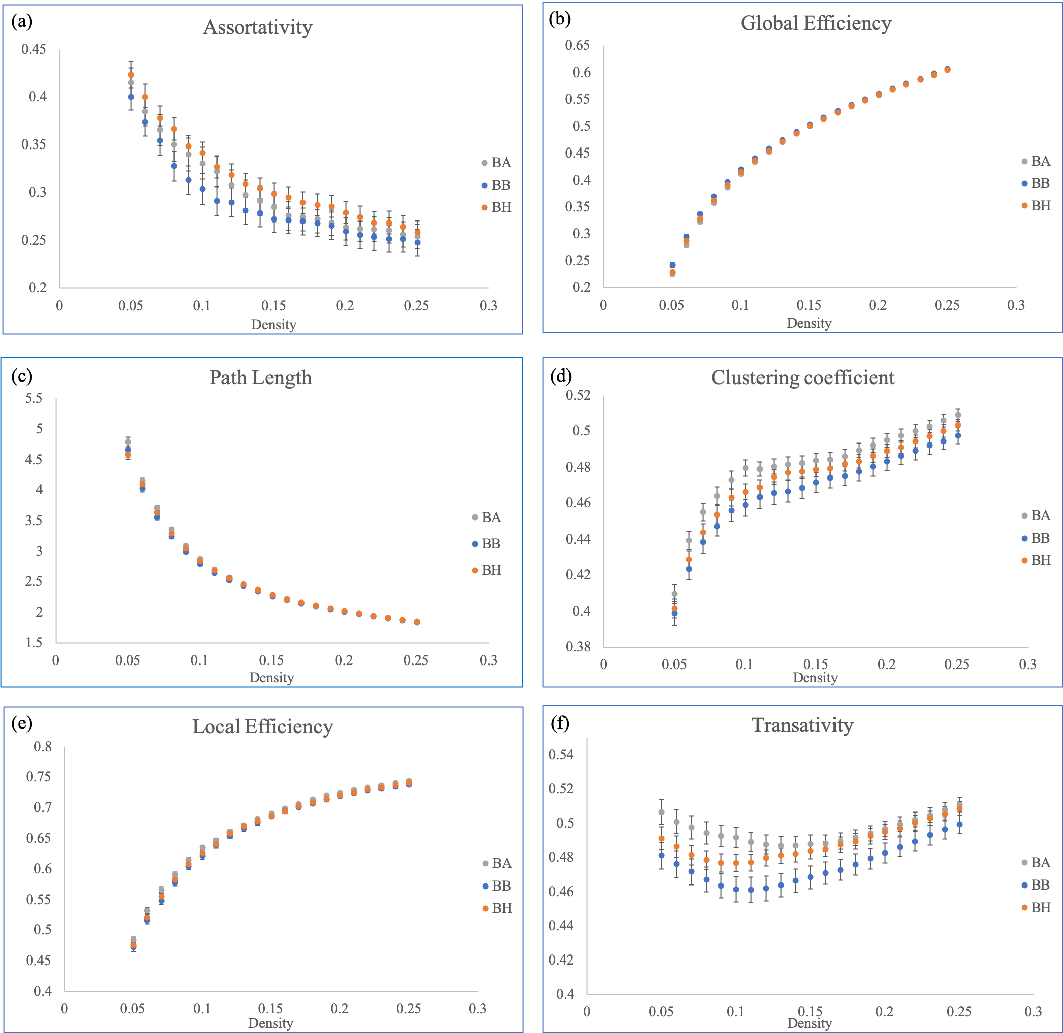
Smoothed fMRI data were employed for graph theoretical analysis. The functional connectivity toolbox, CONN, simplified the connectivity matrix generation procedure. CONN can calculate correlations voxel-to-voxel, but this can be time-consuming. To minimize the possible connections in the voxel space, we first segmented the whole brain of each subject into 90 regions based on the Automated Anatomical Labeling atlas, with each region considered a node. The connection between each node was viewed as an edge. The 90 × 90 connectivity matrix was then computed through Pearson correlations for each participant. Graph Analysis Toolbox (GAT) accepted the output connectivity matrix of each group from CONN. GAT was used to obtain the topological parameters, and the area under the curve (AUC) within chosen ranges for each topology index and comparing them between each group. Topological parameters were conducted in densities between 0.05 and 0.25, with an increment of 0.01. The density represents the ratio of existing connections to possible connections. A two-sample t test and nonparametric permutation test (×1000) was performed by GAT for statistical comparison.
Results
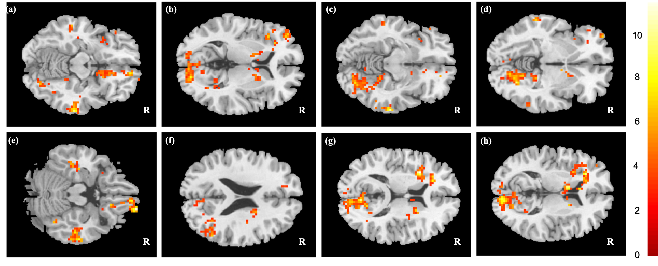
ANOVA performed to determine differences among the three groups revealed significant difference (p < 0.05) in the prefrontal cortex (PFC), bilateral middle, right inferior temporal gyrus, right angular gyrus, left insula, and left caudate (Fig. 1).For graph theoretical analysis, subtle changes in the C+ and C- groups compared with the HC group were demonstrated. Significant AUC comparison were presented in global efficiency, characteristic path length, clustering coefficient, local efficiency, and transativity between C+ and C- group (Fig. 2).
Discussion
Depression severity could be a factor that impacted the result, one of our major finding, caudate, in mfALFF showed statistical significance in ANOVA. The mfALFF represent the cerebral activity of human brain, Fang, et.al. have discovered bilateral caudate reduces glucose metabolism in prechemotherapy patients with depression, as noted under fluorodeoxyglucose positron emission tomography 2.Previous studies suggest that breast cancer and chemotherapy may altered brain network. In our topological results, those changes in the local cliques can also be affected by mood. People diagnosed as having major depressive disorders demonstrated significant decreases in local connections. The alteration of network measures may be associated with memory performance. Shifting network topology in the groups might have been connected to memory performance. Global efficiency, local efficiency, and age may be associated with working memory performance 3; however, this inference warrants further psychological evaluation.
Conclusions
Our study showed subtle alterations of brain activity and networks in cancer survivors, these suggest that functional network disruptions may occurred with and without the administration of chemotherapeutic agent. Further longitudinal and experimental research should be conducted to confirm the mechanisms underlying these alterations.Acknowledgements
This study was supported by the research grants MOST107-2221-E-182-054-MY3, MOST106-2221-E-182-079, and MOST104-2314-B-040-001 from the Ministry of Science and Technology, Taipei, Taiwan. This study was also supported by grants BMRPD1H0102 and BMRPD1G1322 from Chang Gung University, Taoyuan, Taiwan and CORPG6G0102 and CORPG6G0122 from Chang Gung Memorial Hospital, Chiayi, Taiwan.References
1. Hermelink, K., Chemotherapy and Cognitive Function in Breast Cancer Patients: The So-Called Chemo Brain. J Natl Cancer Inst Monogr 2015, 2015 (51), 67-9.
2. Fang, L.; Yao, Z.; An, J.; Chen, X.; Xie, Y.; Zhao, H.; Mao, J.; Liang, W.; Ma, X., Topological Organization of Metabolic Brain Networks in Pre-Chemotherapy Cancer with Depression: A Resting-State PET Study. PLoS One 2016, 11 (11), e0166049.
3. Stanley, M. L.; Simpson, S. L.; Dagenbach, D.; Lyday, R. G.; Burdette, J. H.; Laurienti, P. J., Changes in brain network efficiency and working memory performance in aging. PLoS One 2015, 10 (4), e0123950.
Figures