3997
Surface-Based Morphometry of Brain in Benign Childhood Epilepsy with Centrotemporal Spikes1Medical Imaging, the Affiliated Hospital of Zunyi Medical University, Zunyi, China, 2GE Healthcare,MR Research China, Beijing, China
Synopsis
This study investigated the cortical morphological changes of drug-naïve Benign childhood epilepsy with centrotemporal spikes (BECTS) using three-dimensional T1- weighted images(3D-T1WI) and explored the possible influence of cortical morphological changes on cognition.The results revealed aberrant morphology in thickness, gyrification and fractal dimension, in addition to Rolandic region. Cortical gyrification of right parahippocampal gyrus in BECTS may indirectly reflect the degree of language transmission damage or the result of contralateral compensation.
Introduction
Benign childhood epilepsy with centrotemporal spikes (BECTS),which is self-limited focal epilepsy and also called Rolandic epilepsy,accounts for 8–25% of epilepsy in children. It is characterized by seizures during nocturnal sleep and centrotemporal spikes in electroencephalogram. Some prevenient neuroimaging studies revealed abnormalities of brain structure and cognitive function in patients with BECTS. Considering the influence of antiepileptic drugs and the wide range of patients’ age, the results require further validation. In this study we investigated the cortical morphological changes of drug-naïve BECTS using 3D-T1WI and explored the possible influence of morphological changes on cognition.Methods
The study was approved by the local Institutional Review Board and all the informed written consents were obtained from all parents of participants.Twenty-five drug-naïve BECTS children (age: 9.08 ± 1.55 years, range: 7-13 years,12 males, years in education: 3.00 ±1.35 years,range:1-6 years) and twenty healthy volunteers (age:9.50 ±1.53years,range:7-11years,14 males,years in education:3.60 ±1.60years,range:1-6 years) were enrolled. All participants underwent MR exams (3.0T HDxt, GE Healthcare, Milwaukee, WI) including a 3D-T1 acquisition (TR/TE = 7.8/3.0 ms, TI = 450 ms, FA = 15°, FOV = 256mm×256 mm, matrix = 256x256, slice thickness = 1mm).The neuropsychological assessment based on the Wechsler Intelligence Scale (for children-Chinese Revised) was conducted by an experienced neuropsychologist on the same day of MRI exams. 3D-T1WI data analysis was performed with the CAT12 toolkit, which was based on SPM12 (http://dbm.neuro.uni-jena.de/cat/). Two-sample t tests was carried out to compare the thickness, fractal dimension and gyrification of encephalic regions in DK40 between the patient and control groups. Spearman correlation analysis was uesd to demonstrated the relationships between abnormal encephalic regions and intelligence quotient (IQ) which included verbal intelligence quotient (VIQ), performance intelligence quotient (PIQ) and all intelligence quotient (AIQ). In this study, p< 0.05 was considered statistically significant.Results
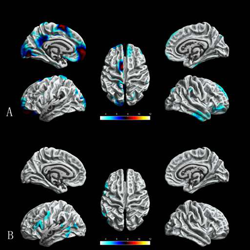
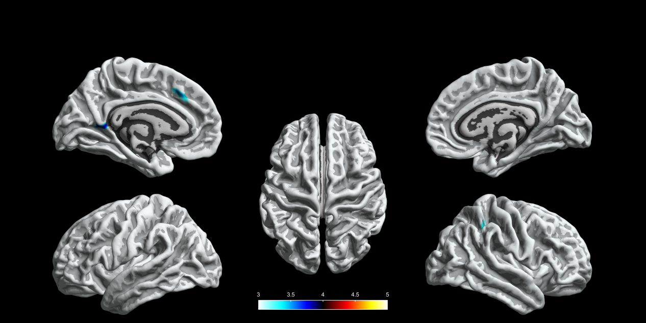
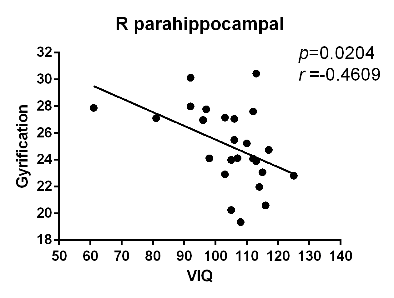
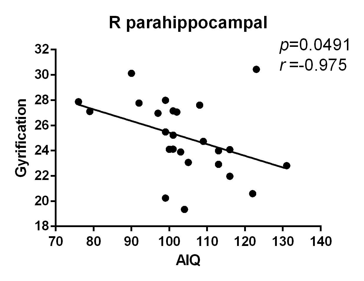
There were no significant differences in age, gender composition, or years in education between the patient group and control group (all p>0.05). The patient group showed extensive cortical thinning predominantly in bilateral frontal, temporal regions and limbic system(Figure1,all p<0.05).In addition, patient group showed significantly increased cortical gyrification in bilateral frontal, parietal, limbic system and right temporal regions, and significantly decreased cortical gyrification in left frontal, occipital, temporal regions and limbic system(Figure2,all p<0.05). Significantly increased fractal dimension was found in left superiorfrontal, caudalmiddlefrontal, bankssts, precuneus and right superiorparirtal gyri (Figure3,all p<0.05)).Correlation analysis revealed that VIQ(Figure 4) and AIQ(Figure5) in BECTS patients were negatively correlated with cortical gyrification in right parahippocampal gyrus(all p<0.05).Discussion and conclusion
BECTS showed aberrant morphology in thickness, gyrification and fractal dimension, mainly in the frontal regions and limbic system.This may provide the underlying mechanism of the cognitive impairment, especially in language and memory function for BECTS patients. Abnormal encephalic regions extended beyond the Rolandic region because epilepsy activity may spread to other cortical areas.Moreover, right parahippocampal gyrus is an important source of afferent fibers in the hippocampus. The relationship between the cortical gyrification changes and VIQ in BECTS may indirectly reflect the degree of language transmission damage or the result of contralateral compensation.Acknowledgements
This work was supported by the National Natural Science Foundation of China (grant No.81901732,81760309), Science and Technology Foundation of Health and Family Planning Commission of Guizhou Province (grant No.gzwjkj2017-1-013),Guizhou Science and Technology Plan Project (grant No.Guizhou Science and Technology platform talents [2017] 5620).
References
[1] Dryzalowk P, Jozwiak S, Franckiewicz M,et al.Neurologia neurochirurgia polska, 2018.
[2] Kim E H, Yum M S, Shim W H, et al. Seizure, 2015,27:40-46.
[3] Fujiwara H, Tenney J, Kadis D S,et al.Acta neurologica Scandinavica, 2018,138(5):432-40.
[4] Baumer F M,Cardon A L, Porterb E. The Journal of pediatrics, 2018,194:13-21.
[5] Kubera K M,Schmitgen M M,Hirjak D,et al.NeuroImage Clinical,2019, 23:101913.
Figures