3985
Altered cerebro-cerebellar effective connectivity in new-onset juvenile myoclonic epilepsy1Lanzhou University Second Hospital, Lanzhou, China
Synopsis
Resting-state fMRI studies have indicated that juvenile myoclonic epilepsy (JME) could cause widely functional connectivity disruptions between cerebrum and cerebellum. However, the directed influences or effective connectivities (ECs) bewteen these brain regions are poorly understood. In the current study, we aim to evaluate the ECs between cerebrum and cerebellum in new-onset patients with JME.
Purposes
Purposes: Juvenile myoclonic epilepsy (JME) is a common idiopathic generalized epilepsy (IGE) syndrome characterized by impairments in executive and cognitive control, affecting independent living and psychosocial functioning. There is a growing consensus that JME is associated with abnormal function of diffuse brain networks, typically affecting frontal and fronto-thalamic areas,however whether the cerebellum participate the networks are poorly understood.We aimed to evaluate structural and functional connectivity of patients with newly diagnosed juvenile myoclonic epilepsy (JME) compared to healthy subjects.Methods
Methods:Data were collected on a MAGNETOM Verio 3T MR scanner (SiemensHealthcare, Erlangen, Germany) with a 8-channel head coil.We enrolled 34 patients with newly diagnosed JME and age-, sex-, education- matched HCs (n = 34) participated in the current study and underwent the rs-fMRI scanning. We used degree centrality (DC) to investigate functional connectivity (FC) strength of the whole-brain network and Granger causality to analyze effective connectivity in order to explore directional aspects involved in JME.Results
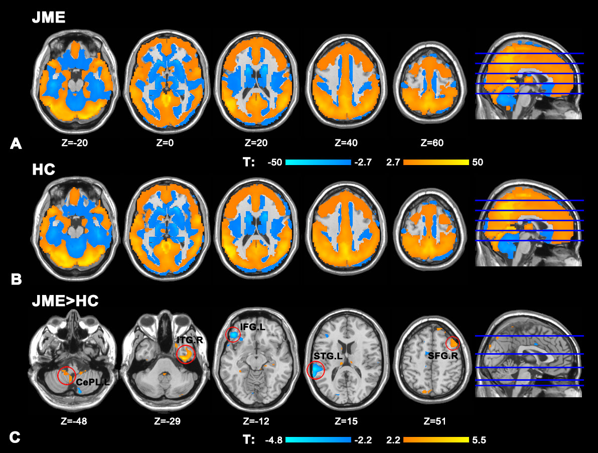
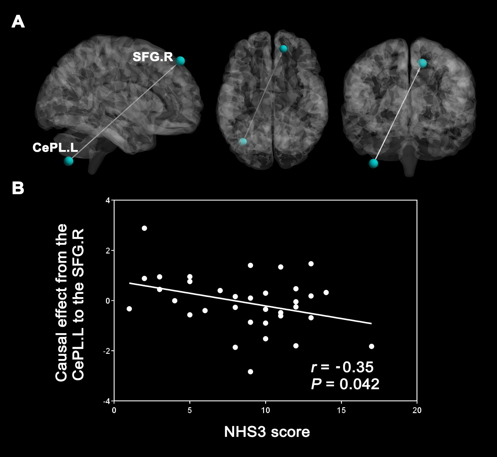
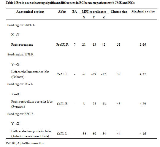
Results:Compared to HCs, we found significantly increased DC in Left cerebellum posterior lobe (CePL.L)、Right inferior temporal gyrus(ITG.R)and Right superior frontal gyrus (SFG.R)and decreased DC in Left inferior frontal gyrus(IFG.L)and Left superior temporal gyrus(STG.L). Unidirectionally, CePL.L revealed increased effective connectivity to the Right precuneus(PreCU.R),while the ITG.R revealed decreased effective connectivity to the Left cerebellum anterior lobe(CeAL.L),the IFG.L decreased effective connectivity to the Right cerebellum posterior lobe(CePL.R),and the SFG.R decreased effective connectivity to the Left cerebellum posterior lobe(CePL.L).In addition,the effective connectivity from the CePL.L to the SFG.R showed positive correlations with HNS3 score.Conclusions
Conclusions: Rs-fMRI provides a new and novel method for identifying aberrant brain network architecture. JME patients have disrupted FC strength and causal connectivity mostly in non-motor regions, especially the anterior and posterior lobes of the cerebellum. The current findings will provide a new perspective for understanding the neuropathophysiological mechanisms in JME.Acknowledgements
This study was supported by Cuiying Scientific and Technological Innovation Program of Lanzhou University Second Hospital.References
1.Chen YC, Feng Y, Xu JJ, et al.Disrupted Brain Functional Network Architecture in Chronic Tinnitus Patients.Front Aging Neurosci. 2016 Jul 8;8:174.
2.Jiang W, Lei Y, Wei J, et al.Alterations of Interhemispheric Functional Connectivity and Degree Centrality in Cervical Dystonia: A Resting-State fMRI Study.Neural Plast. 2019 Apr 24;2019:7349894.
3.Wang Y, Berglund IS, Uppman,et al.Juvenile myoclonic epilepsy has hyper dynamic functional connectivity in the dorsolateral frontal cortex.Neuroimage Clin. 2019;21:101604.
4.Zhang Z, Liu G, Yao Z,et al.Changes in Dynamics Within and Between Resting-State Subnetworks in Juvenile Myoclonic Epilepsy Occur at Multiple Frequency Bands.Front Neurol. 2018 Jun 14;9:448.
5.Zhong C, Liu R, Luo C,et al.Altered Structural and Functional Connectivity of Juvenile Myoclonic Epilepsy: An fMRI Study.Neural Plast. 2018 Feb 26;2018:7392187.
Figures