3984
The effect of meditation on brain functional connectivity using dynamic ASL1Computer Science, State University of New York at Binghamton, Binghamton, NY, United States, 2National Institute on Aging, National Institutes of Health, Baltimore, MD, United States
Synopsis
We applied dynamic arterial spin labeling (ASL) in detecting the changes of brain functional connectivity (FC) in ten college students after 2-months of meditation practice. FC maps before and after meditation practice were compared, and the changes of FC were correlated to practice time. We observed that the increased practice time was associated with increased FC between long-distance nodes within default mode network (DMN) and within dorsal attention network (DAN), between DMN and salience network (SN), with decreased FC between short-distance nodes within DMN and DAN. The findings support efficient global communication even after a short-term meditation for two months.
Introduction
Resting-state BOLD fMRI has shown the functional connectivity (FC) changes of brain networks in default mode network (DMN) and dorsal attention network (DAN) during the meditation practice [1, 2]. It is not clear whether the changes in brain networks can be sustained during the rest period, even when meditation is not being performed, for its potential benefits in a clinical population. Dynamic arterial spin labeling (dASL) has demonstrated its capability in detecting resting-state networks [3, 4] and its insusceptibility to physiological noises. It is interesting to ask whether the changes in brain networks can be sustained during the rest period, even when meditation is not being performed, for its potential benefits in a clinical population. Here, we explore whether dASL can detect the sustained change in brain FC during rest following 2-months of meditation practice by a longitudinal design.Methods
All studies were conducted using a GE 3T MR 750. Volunteers received 3D T1-weighted images for image registration and dynamic pseudo-continuous arterial spin labeling (PCASL) sequence [3] for FC measurements before and after 2-months of meditation practice (practice duration: 66.504.14 days, practice time: 574.00312.30 minutes). Ten college students (19.200.28 years old, age range: 19 to 20 years old, 4 females) were recruited from a university meditation class. Subjects were instructed to practice focused meditation for a minimum of 10 minutes each time and at least 5 times per week. They were allowed to choose their own focus: their breath, a point on the wall, a phrase, or anything else as they saw fit. At the end of follow-up scans, volunteers reported their practice time. For each subject, the ASL time series were transformed to the standard brain space using the T1-weighted images as an intermediate. The global mean signals were regressed out from the ASL image time series. Five seed regions of interest (ROIs) [5] were chosen from DMN and DAN: the posterior cingulate cortex(PCC), left visual (LV), right visual (RV), left superior parietal area (LSP) and right superior parietal (RSP)areas. Individual FC maps were calculated from global signal regressed ASL time series using Pearson correlation between time series from the seed ROIs and those from individual voxels throughout the entire brain. Individual FC maps were transformed into z score maps by using a Fisher z transformation to improve normality for group level t tests and smoothed with a 6×6×6 mm Gaussian kernel. The z score maps before and after meditation practice were compared using SPM8 via a paired t test with gender as a covariate. Age was not considered as a covariate because the maximum difference among ages of our subject is 1 year. The difference of the z score maps before and after practice meditation was modeled using SPM8 via multiple linear regression with gender and practice time as covariates. The statistical maps were thresholded using a voxel-level p value of 0.01. A cluster-level p value of 0.05 was used to correct for multiple comparisons. Regional analysis has been used to verify the correlation of the FC changes with practice time. Regional FC values at the clusters which showed significance in the voxel-based analysis were calculated both at the baseline and follow-up. The change of regional FC values was modeled via a linear regression model with gender and practice time as variables.Results & Discussion
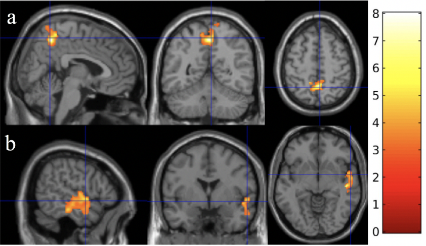
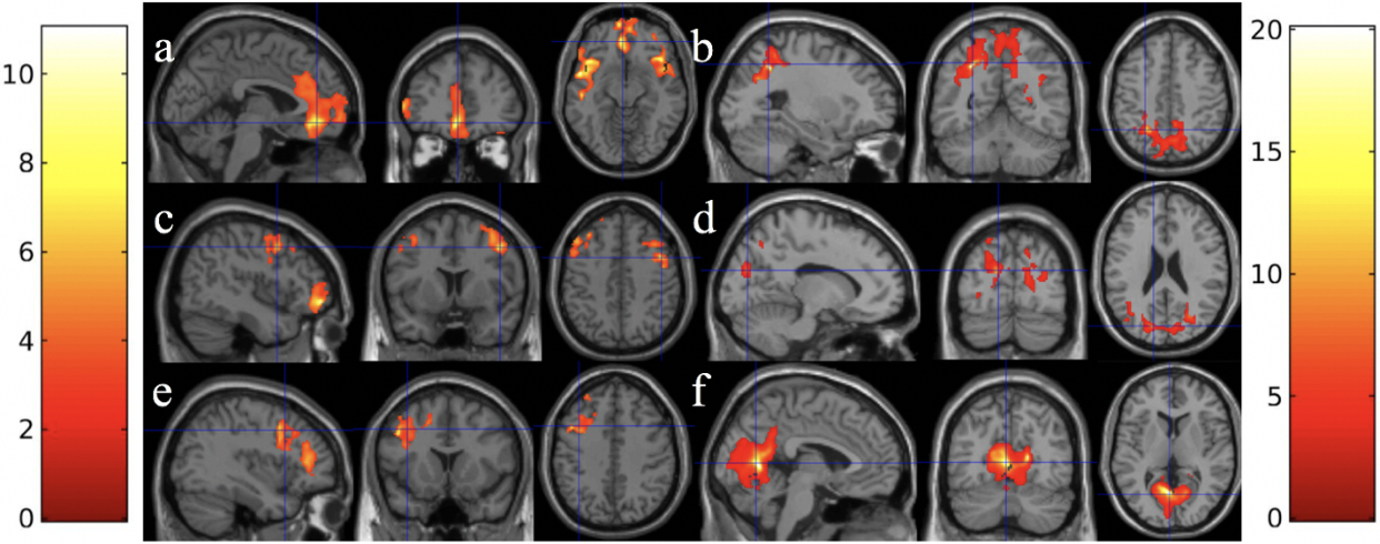
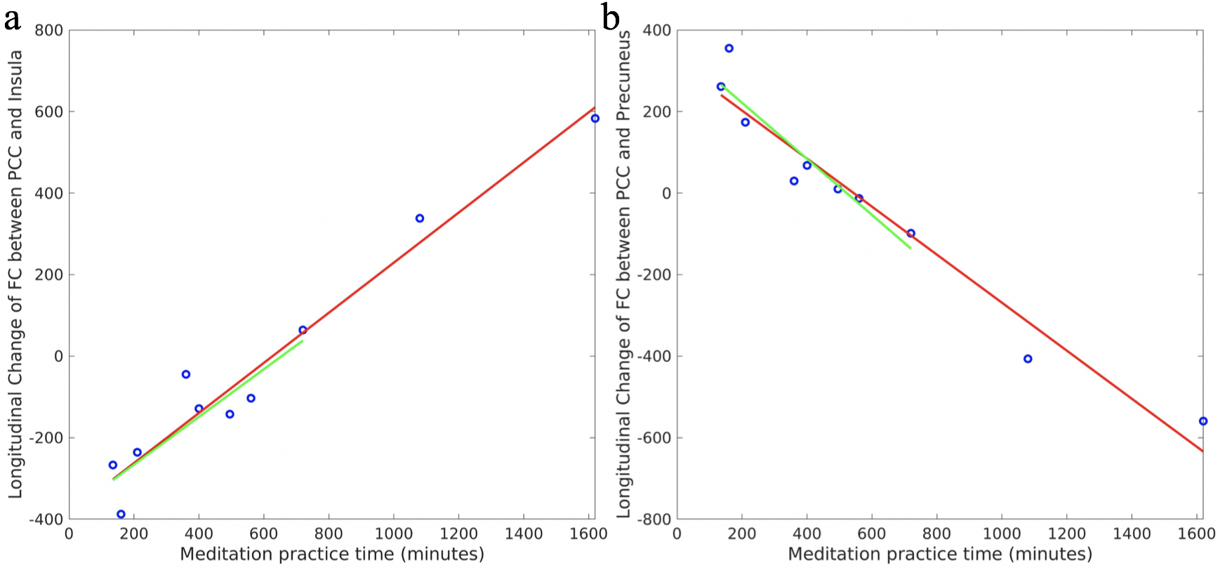
We observed significantly increased FC between the LV seed and precuneus area (Fig. 1a), and decreased FC between the LV seed and superior temporal area (Fig. 1b) after 2-months of meditation practice. Meditation practice time was positively associated with FC between the PCC seed and insular and prefrontal areas (Fig. 2a), between the RV seed and mid frontal area (Fig. 2c), and between the LSP seed and mid frontal area (Fig. 2e). This indicates that the meditation can render the FC of long-distance nodes (regardless whether they belong to the same network) stronger. Meditation practice time was negatively associated with FC between the PCC seed and precuneus area (Fig. 2b), between the RV seed and occipital area (Fig. 2d), and between the LSP seed and occipital area (Fig. 2f). This suggests that the meditation can render the FC of short-distance nodes (nearby regions) weaker. Post-hoc regional analysis showed that the longitudinal change of FC may change from negative to positive (Fig. 3a) or from positive to negative (Fig. 3b) as more meditation practice time is involved.Conclusion
Short-term meditation can increase the communication for the long-distance nodes within DMN and within DAN, between DMN and salience network (SN), supporting efficient global communication even after a short-term meditation.Acknowledgements
No acknowledgement found.References
1. Kilpatrick, L.A., et al., Impact of Mindfulness-Based Stress Reduction training on intrinsic brain connectivity. Neuroimage, 2011. 56(1): p. 290-8.
2. Josipovic, Z., et al., Influence of meditation on anti-correlated networks in the brain. Front Hum Neurosci, 2011. 5: p. 183.
3. Dai, W., et al., Quantifying fluctuations of resting state networks using arterial spin labeling perfusion MRI. J Cereb Blood Flow Metab, 2016. 36(3): p. 463-73.
4. Zhao, L., et al., Global Fluctuations of Cerebral Blood Flow Indicate a Global Brain Network Independent of Systemic Factors. J Cereb Blood Flow Metab (In Press), 2017.
5. Vincent, J.L., et al., Evidence for a frontoparietal control system revealed by intrinsic functional connectivity. J Neurophysiol, 2008. 100(6): p. 3328-42.
Figures