3983
Study on brain function changes in patients with neurological depression based on resting state functional magnetic resonance imaging(rs-fMRI)1Jilin University First Hospital, Changchun, China, 2Philips healthcare, Guangzhou, China
Synopsis
The global incidence of depression is gradually increasing nowadays. However, the exact pathogenesis mechanism is not very clear. Traditional diagnosis approaches to depression usually rely on clinical scales, which is sometimes even subjective. It is essential to make up objective and repetitive methods to diagnose the depression. Resting-state functional magnetic resonance imaging (rs-fMRI) offers a noninvasive technique for detecting and measuring subtle functional brain activities of specific regions. This study aims to explore the change of brain function area in patients with depression and assess the role of these altered brain regions in the pathogenesis.
Purpose
To construct functional brain network using rs-fMRI approach and detect the changes of brain functional connectivity in patients with depression, and to compare the relationship between rs-fMRI changes and clinical scales in evaluating the depression. To provide imaging evidence for the diagnosis and pathogenesis of patients with depression.Material and Methods
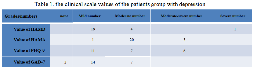
This study included 24 patients with depression scored by 24 Hamilton Depression Scale (HAMD) and 16 healthy controls(HC) matched for age, sex and education level. Firstly, all subjects were inquired medical history characteristics by professionally trained neurologists. Hamilton Anxiety Scale (HAMA), 24 HAMD, Patient Health Questionnaire Depression Scale (PHQ-9), General Anxiety Scale (GAD-7) were scored(IntelliSpace Discovery, Philips, Best, The Netherlands). Secondly, all subjects were collected for rs-fMRI data; then brain functional networks were constructed based on the fractional amplitude of frequency fluctuation(fALFF), regional homogeneity(ReHo), and functional connectivity(FC) data. At last, different brain regions and abnormal functional connectivity characteristics were determined by comparing fALFF, ReHo, and FC data of the two groups. Image preprocessing uses DPARSFA (Data processing assistant for resting-state fMRI http://www.restfmri.net/forum) software based on Matlab (R2018b) platform. SPSS 24.0 software was used for statistical analysis of general data, and a two-sample T-test was performed to compare the general data and clinical evaluation between the two groups. The fALFF data, ReHo data, and FC data of the two groups were subjected to a one-sample T-test by using dpadi software based on Matlab (R2018b) platform (http://rfmri.org/dpabi) and SPM12 software (Statistical parametric mapping http://www.fil.ion.ucl.ac.uk/spm/) respectively. Subsequently, a two-sample T test between groups was performed to calculate the fALFF value, ReHo value, and FC value of the two groups, and P < 0.01 was considered to be statistically significant. In the end, the two groups were compared by the calculated values of areas of fALFF, ReHo, and FC to find out which areas were enhanced or weakened.Results
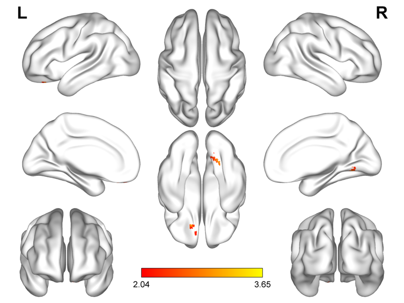
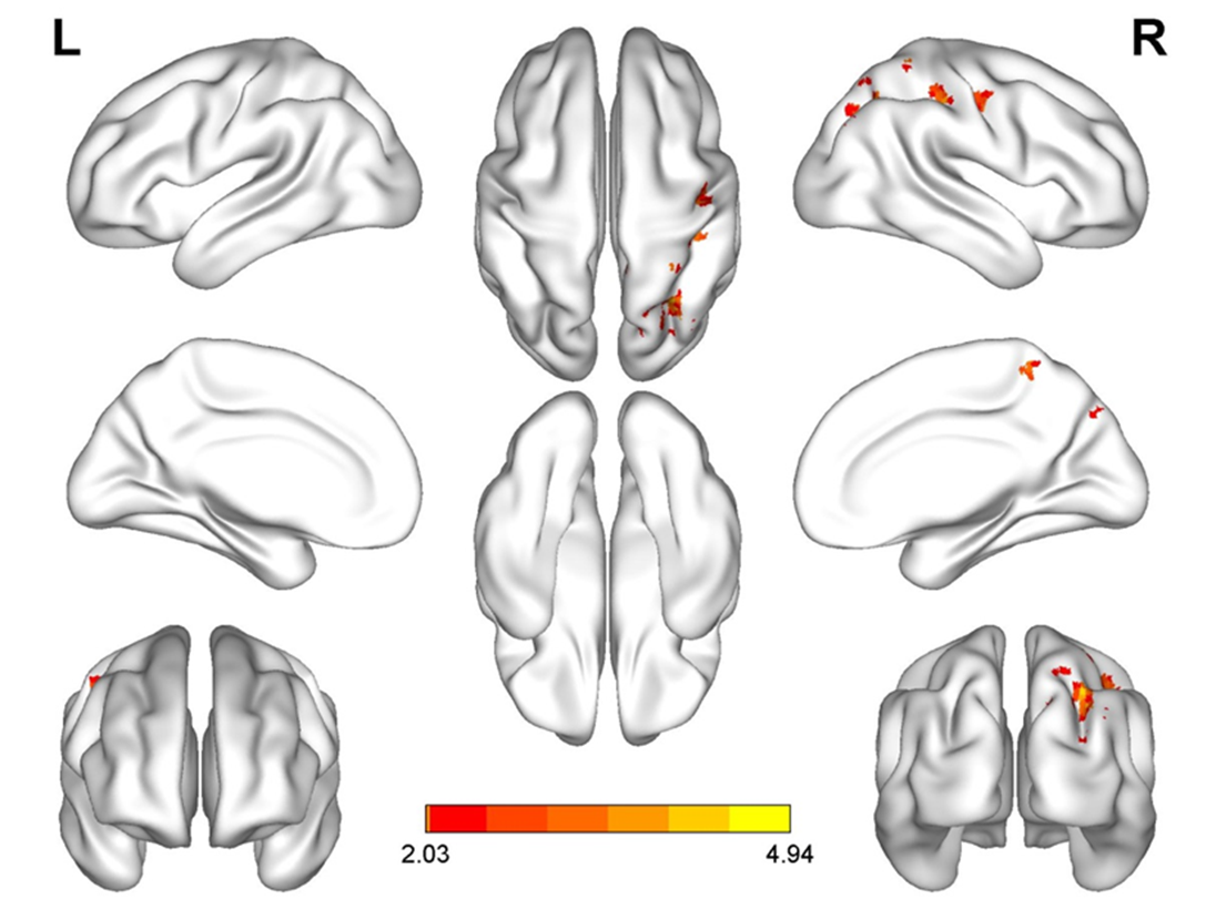
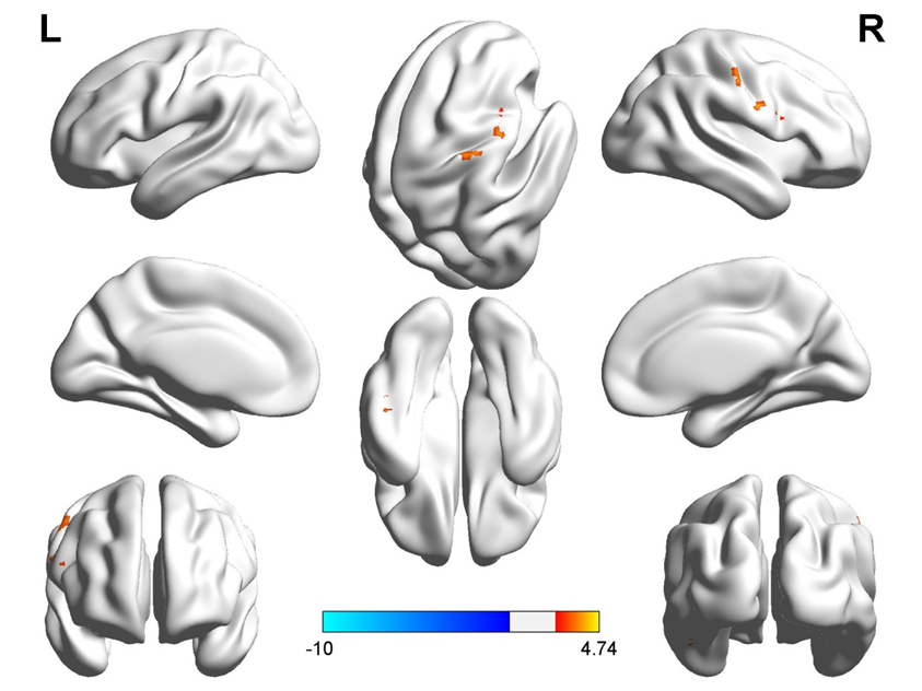
There was no significant difference in gender, age, occupation and education level between the patient group with depression and the healthy control group (P>0.05). There was a significant difference in HAMD score, HAMA score, PHQ-9 score and GAD-7 score between the two groups (P<0.05)(Tab.1). The patients with physical symptoms as the primary complaint accounted for about 83%, and the emotional symptoms were the first complaints accounting for 17%. Common physical symptoms were mainly caused by sleep disorders, dizziness, headache, memory loss, etc. The rs-fMRI images of the patient group and the healthy control group: the fALFF values in the left superior frontal gyrus, the right cerebellum 6 area and the gyrus lingualis were higher in the patient group than those of the control group(Fig.1); The ReHo value in the right posterior central gyrus, the right superior occipital lobe and the right angular convolution was higher in the patient group than those of the normal control group(Fig.2); Compared with those of the control group, there is an increase of FC values of patient group in the right posterior central gyrus, the right gyrus temporalis medius, the left fusiform gyrus, the left inferior temporal gyrus and the medial prefrontal cortex (Fig.3).Discussion
The number of women with depression in neurology clinics is higher than males, mostly young and middle-aged1. The study found that there are much activation areas difference between the control and the depression. The fALFF values in the left superior frontal gyrus, the right cerebellum 6 area and the gyrus lingualis were higher in the patient group, which meant the local neurons in the above area increasing their activity2. The ReHo value in the right posterior central gyrus, the right superior occipital lobe and the right angular convolution was higher. The synchronization of local neuronal activities in the above region was enhanced3. Compared with the control group, the FC values of the patient group in the right posterior central gyrus, the right gyrus temporalis medius, the left fusiform gyrus, the left inferior temporal gyrus and the medial prefrontal cortex were increased, which means that the functional connection between the upper region and the medial prefrontal lobe enhanced4. Changes in the function of these sites may play an essential role in the pathogenesis of depression, henceforth provide new evidence for the pathogenesis of depression.Conclusion
By comparing the value changes of fALFF, ReHo, and FC in the healthy control group with those of the depression patients, we could observe a significant number of differences in rs-fMRI between the above groups. The method is very benenificial for the clinician to find out the pathogenesis of depression, the mechanism of the depression and further evaluation of the efficacy of the anti-depression drug.Acknowledgements
No acknowledgement found.References
1.Biller J, Sacco RL, Albuquerque FC, et al. Cervical arterial dissections and association with cervical manipulative therapy: A statement for healthcare professionals from the american heart association/american stroke association[J]. Stroke; a journal of cerebral circulation. 2014.45:3155-3174.
2. Puskas S.Quantitative EEG in obstructive sleep apnea syndrome: a review of the literature.2017,28(3):265-70.
3. Depping MS, Wolf ND, Vasic N, Sosic-Vasic Z, Schmitgen MM, Sambataro F, et al.Aberrant resting-state cerebellar blood flow in major depression[J].J Affect Disord.2018,226:227-31.
4. Yamamura T, Okamoto Y, Okada G, Takaishi Y, Takamura M, Mantani A, et al.Association of thalamic hyperactivity with treatment-resistant depression and poor response in early treatment for major depression: a resting-state fMRI study using fractional amplitude of low-frequency fluctuations[J].Transl Psychiatry.2016,6:e754.
Figures