3974
Resting-state Functional Connectivity during Voluntary Mouth Breathing
Chan-A Park1, Ju-Yeon Jung2, Yeong-Bae Lee3,4, and Chang-Ki Kang2,4,5
1Biomedical Engineering Research Center, Gachon University, Incheon, Korea, Republic of, 2Department of Healthy Science, Gachon University Graduate School, Incheon, Korea, Republic of, 3Department of Neurology, Gachon University Gil Medical Center, Incheon, Korea, Republic of, 4Neuroscience Research Institute, Gachon University, Incheon, Korea, Republic of, 5Department of Radiological Science, Gachon University, Incheon, Korea, Republic of
1Biomedical Engineering Research Center, Gachon University, Incheon, Korea, Republic of, 2Department of Healthy Science, Gachon University Graduate School, Incheon, Korea, Republic of, 3Department of Neurology, Gachon University Gil Medical Center, Incheon, Korea, Republic of, 4Neuroscience Research Institute, Gachon University, Incheon, Korea, Republic of, 5Department of Radiological Science, Gachon University, Incheon, Korea, Republic of
Synopsis
The purpose of the study is to examine the difference of the functional connectivity between the nasal and mouth breathing conditions in healthy subjects using resting-state functional magnetic resonance imaging via seed-based correlation analysis. “Mouth>Nose” contrast had 5 seeds and 23 connecting pairs, however, 6 seeds and 14 pairs in the “Mouth<Nose” contrast. Especially, caudate had the most number of connections of salience networks, supramarginal gyrus, insular cortex, central opercular cortex, supramarginal gyrus, and parietal operculum in “Mouth>Nose” contrast. These indicated that the limbic system regulates the resting-state functional connectivity during the voluntary mouth breathing compared the nasal breathing.
Introduction
Pathological or habitual mouth breathing leads to face deformation, a higher prevalence of asthma, and low academic achievements in children. 1,2 Although various side effects are associated with such a condition, fundamental research on the changes in brain activity caused by mouth breathing has hardly been conducted. 3 Furthermore, there is little investigation about alterations of the resting-state functional connectivity (FC) in the whole-brain regions in the voluntary mouth breathing condition using the resting-state functional magnetic resonance imaging (fMRI). The aim of the study is to examine the difference of the whole-brain FC network between mouth and nasal breathing conditions in the resting-state fMRI via seed-based correlation analysis.Methods
Twenty-three healthy subjects (male:female = 10:13; age = 22.43 ± 1.34 years) participated in the study and provided written informed consent. The study protocol was approved by the institutional review board (IRB). The experiment was performed using a 3T magnetic resonance imaging (MRI) scanner (Siemens Verio, Erlangen, Germany) with a commercially available 12-channel radio-frequency (RF) head matrix coil. All participants underwent two imaging sequences in MRI, that is, a high-spatial-resolution T1-weighted anatomical imaging sequence of three-dimensional (3D) magnetization–prepared rapid acquisition gradient echo (MP-RAGE); and a blood-oxygen-level-dependent (BOLD) fMRI sequence of two-dimensional (2D) echo planar imaging (EPI) with repetition time (TR)/echo time (TE) = 2000 ms/30 ms, and flip angle (FA) = 90°. The participants were asked to breathe voluntarily only through their mouth or nose at the beginning of each session. Subjects were instructed not to move their head as much as possible with eyes closed and stay awake throughout the scanning. Each subject put nose plug on their nostrils during mouth breathing condition in order to prevent nose breathing, whereas they took off the nose plug during nasal breathing condition to induce a natural nasal breathing with the mouth closed. The CONN (version 18b) FC toolbox (www.nitrc.org/projects/conn) is a MATLAB-based software for the calculation, display and analysis of resting-state FC in resting-state fMRI studies. The CONN was used to calculate the strength and significance of the bivariate correlation among region of interest (ROI) pairs within all subjects’ data in the “Nose” and “Mouth” breathing conditions. To measure the level of linear association of the BOLD time series, a bivariate correlation was used to conduct the first-level analysis, in which the effect size is the correlation coefficient. 4 In ROI-to-ROI analysis, we tested both “Mouth>Nose” and “Mouth<Nose” contrasts to create connections between whole ROIs and to perform networks analysis with one-sided inferences. The connectome ring maps were constructed as the thresholded intensity of ROI-to-ROI connection with one-sided positive seed level correction and permutation tests. The significance of ROI-to-ROI connection was determined through the false discovery rate (FDR)-corrected P < 0.05 with seed-level correction.Results
ROI-to-ROI FC connectome ring (Fig. 1) showed more connections in the “Mouth” breathing condition than the “Nose” breathing condition. The “Mouth>Nose” contrast had significantly stronger connections in 19 ROIs (Fig. 2a) and 5 ROI-to-ROI networks (Fig. 3a). Among them, the left and right lateral sensorimotor area had the most significant connection (P < 0.05, T = 5.67), and the left caudate had the biggest intensity value (P < 0.05, Intensity = 35.96) and 10 connected seeds which had the most number of connection pairs between other seeds including salience networks (anterior cingulate cortex and supramarginal gyrus), right supramarginal gyrus with posterior division, left and right insular cortex, left central opercular cortex and supramarginal gyrus with anterior division, and parietal operculum (P < 0.05, Size = 10). However, the “Mouth<Nose” contrast had 10 significant stronger ROI-to-ROI connections (Fig. 2b) and 6 networks (Fig. 3b). Among them, the left lateral sensorimotor area and superior sensorimotor area had the most significant connection (P < 0.05, T = 6.86). The superior sensorimotor area had the biggest intensity value (P < 0.05, Intensity = 33.89) and 7 connected seeds which had the most number of connection pairs between other seeds including the left and right lateral sensorimotor area and precentral gyrus, left postcentral gyrus, left and right central opercular cortex (P < 0.05, Size = 7).Discussion
The results showed that when compared with the nasal breathing condition, the mouth breathing condition showed higher functional connections in the whole-brain ROIs. Among them, basal ganglia, especially left caudate, exhibited the most number of connections with other regions in the “Mouth>Nose” contrast. According to the previous study, the caudate has a significant relation with the respiratory sensation and motor process. These indicated that the limbic system could regulate the resting-state functional connectivity during the voluntary mouth breathing compared to the nasal breathing in healthy subjects.Conclusion
In the present study, we demonstrated that the resting-state FC during voluntary mouth breathing condition could significantly induce different connections of ROIs compared to during nasal breathing condition in healthy subjects. These findings suggested that FC of the voluntary mouth respiration was much more associated with mouth sensation and motor process.Acknowledgements
This research was supported by Basic Science Research Program through the National Research Foundation of Korea (NRF) funded by the Ministry of Education (grant number: NRF-2019R1I1A1A01058253).References
- Izu SC, Itamoto CH, Pradella-Hallinan M et al. Obstructive sleep apnea syndrome (OSAS) in mouth breathing children. Braz J Otorhinolaryngol. 2010;76(5):552-556.
- Jefferson Y. Mouth breathing: adverse effects on facial growth, health, academics, and behavior. Gen Dent. 2010;58(1):18-25.
- Park CA, Kang CK. Sensing the effects of mouth breathing by using 3-tesla MRI. J Korean Phys Soc. 2017;70(12):1070-1076.
- Whitfield-Gabrieli S, Nieto-Castanon A. Conn: a functional connectivity toolbox for correlated and anticorrelated brain networks. Brain Connect. 2012;2(3):125-141.
Figures
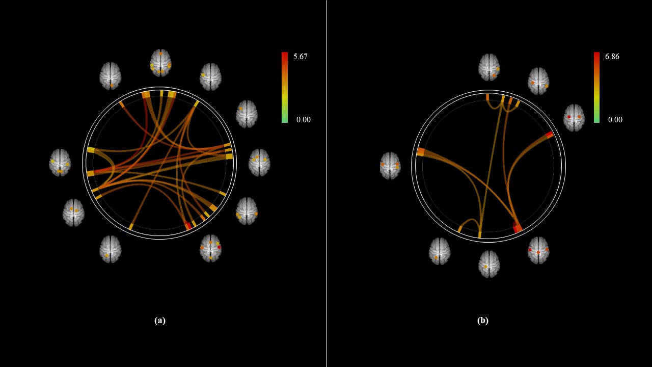
Fig. 1. ROI-to-ROI connectome ring maps of all
selected ROI seeds in “Mouth>Nose” (a) and “Mouth<Nose” (b) contrasts.
The rings are obtained at the height threshold FDR of P < 0.05 with
one-sided (positive) seed level correction and permutation tests. The color bar
indicates the statistical T value. Abbreviations: ROI, region of
interest; FDR, false discovery rate.
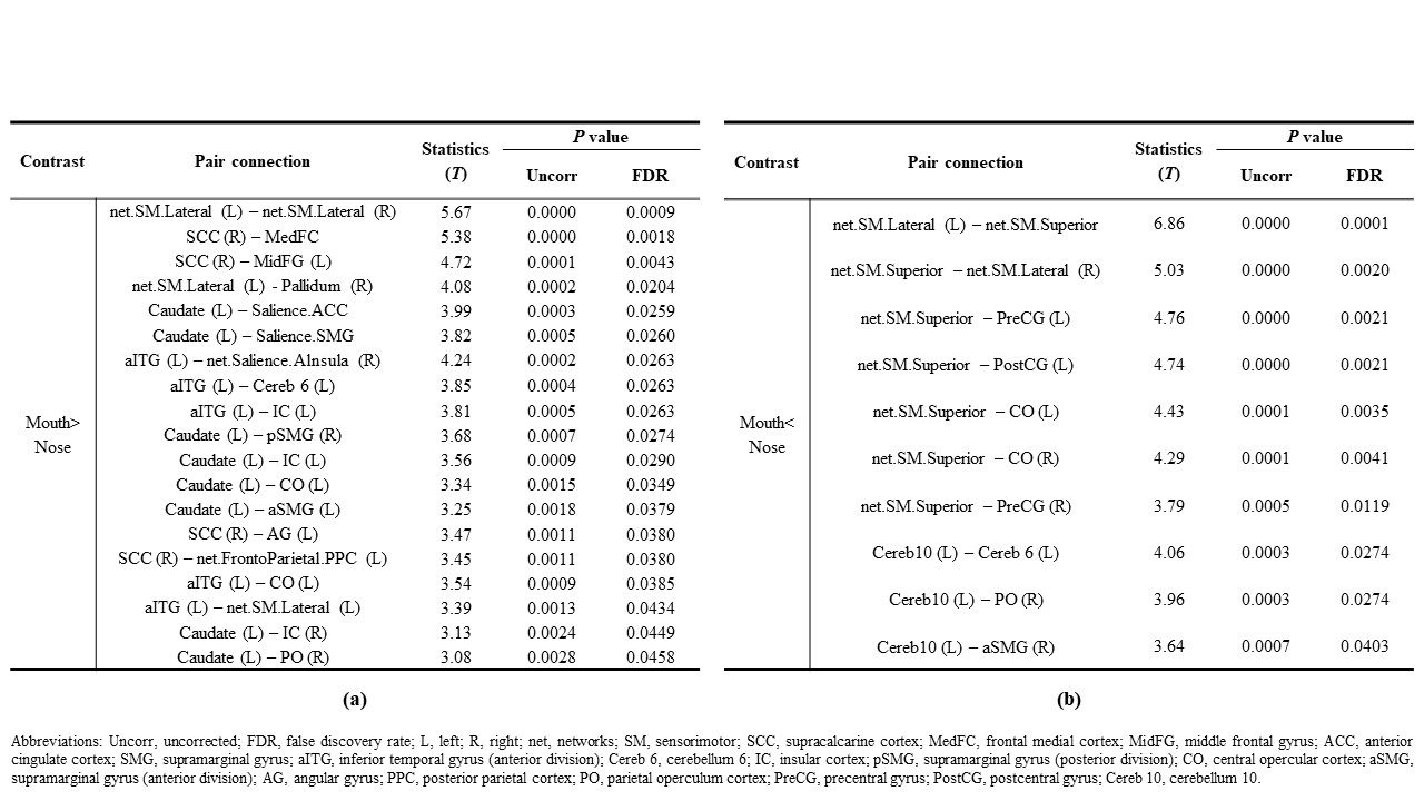
Fig. 2. The
ROI pair connections at a 0.05 one-sided (positive) FDR-p value in (a) “Mouth>Nose” and (b) “Mouth<Nose” contrasts. Abbreviations:
ROI, region of interest; FDR, false discovery rate.
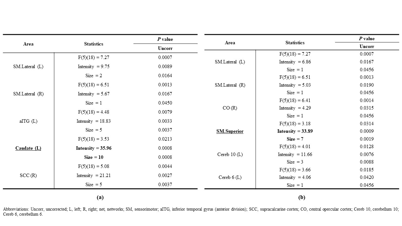
Fig.
3. The connected regions included ROI that were
more significantly connected at a 0.05 one-sided (positive) uncorrected-p value
in (a) “Mouth>Nose” and (b) “Mouth<Nose” contrasts. Abbreviations:
ROI, region of interest.